Small Brown Mushrooms Identification Guide 2026

Nathan Cole
Mycologist · Author · Fungi Expert
Updated
Apr 22, 2026
Small Brown Mushrooms: The Complete Field Guide to LBM Identification, Deadly Lookalikes, and Forager Safety
Small brown mushrooms — mycologists call them LBMs, Little Brown Mushrooms — are the most dangerous category in amateur foraging. Not because any single species is exotic or rare, but because dozens of them occupy the same habitats, look nearly identical in the field, and span the full spectrum from deadly to edible to legally controlled. Galerina marginata kills with the same amatoxins as the death cap.
Cortinarius rubellus destroys kidneys two to three weeks after a single meal. Both grow within feet of species people actively hunt. If you take one thing from this guide: never eat an unidentified small brown mushroom. That's the answer. Everything below is the evidence.
What Makes a Mushroom an "LBM"?
Defining the Little Brown Mushroom Problem
The term "Little Brown Mushroom" isn't a formal taxonomic category. It's a field mycologist's honest admission that a significant portion of the fungal kingdom simply refuses to be easily named. David Arora, in Mushrooms Demystified, practically threw up his hands at this group — dedicating pages to warning readers that LBMs constitute a "bewildering array" of small, brown, unremarkable-looking fruiting bodies that can defeat even experienced collectors.
What qualifies a mushroom as an LBM? Informally: a pileus ranging from tan to tawny to dark umber-brown, typically under 6 cm in diameter, on a slender stipe, often without distinctive features that pop in the field. The group cuts across multiple unrelated genera — Galerina, Cortinarius, Inocybe, Hebeloma, Pholiota, Naucoria, Tubaria, Crepidotus, Gymnopus, Phaeocollybia, Pholiotina — meaning there's no single taxonomic family to anchor your intuition. You're dealing with convergent evolution: unrelated lineages that arrived at the same general shape because it works.
The pileus of most LBMs is hygrophanous — it changes color dramatically as it absorbs or loses moisture. A cap that's deep chestnut-brown after rain may look pale ochre by afternoon the same day. That single feature wrecks most field ID attempts, because color is the first thing novice foragers key on, and color is precisely what you can't rely on here.
Why Even Experts Struggle?
I've been doing this for over 40 years. I've formally identified more than 400 species across the Pacific Northwest, Appalachia, the Gulf Coast, and Western Europe. I still send LBM specimens to the lab.
Here's why: the characters that actually separate genera and species in this group — cheilocystidia morphology, spore ornamentation under Melzer's reagent, clamp connections at hyphal septa, chrysocystidia presence in KOH — are microscopic. You cannot see them in the field. You need a compound microscope, slide-mounted material, and in many cases ITS DNA sequencing to make a definitive call. "I looked it up in a field guide" is simply not adequate for this group.
Even field guides written by excellent mycologists acknowledge the problem. McKnight and McKnight's Peterson Field Guide to Mushrooms of North America explicitly flags entire genera as "not recommended" without microscopic examination. Breitenbach and Kränzlin's Fungi of Switzerland, volumes 4 and 5 — among the most rigorous taxonomic treatments of agarics ever published — hedge on certain Cortinarius and Inocybe complexes.
The NAMA Toxicology Committee has documented poisoning cases involving LBMs across nearly every decade since the 1970s. The pattern is consistent: a forager confidently identifies something based on gross field characters, eats it, and presents to an emergency room days later. By then, with amatoxin ingestion, the damage is done.
That's not a beginner problem. That's a structural problem with this group of fungi. Respect it accordingly.
The Deadly Ones You Must Know First
This section comes before edibility discussions, before habitat guides, before anything else — because the order matters. These species need to be burned into memory before you touch a single small brown mushroom in the field.
Galerina marginata — The #1 Killer in This Group
Galerina marginata is the most dangerous mushroom most foragers have never seriously studied. Amanita phalloides gets the headlines. But Galerina marginata is responsible for a substantial share of fatal amatoxin poisonings in North America — because it grows in urban parks, on wood chip mulch beds, on dead conifer logs in backyards, and it's virtually indistinguishable from species people actively hunt.
Physical description:
- Pileus: 1–5 cm, convex to broadly umbonate, tawny-brown to honey-brown, strongly hygrophanous — fading pale buff from the center outward as it dries
- Stipe: 3–7 cm tall, slender, with a fragile fibrous annulus that frequently disappears with age or rain — the most critical field feature, and also the most unreliable one
- Lamellae: Adnate to slightly decurrent, close, pallid to rusty-cinnamon
- Spore print: Rust-brown to cinnamon
- Habitat: Almost always on wood — dead conifer logs, buried roots, wood chip mulch; occasionally appearing terrestrial when buried substrate isn't visible
- Season: Spring through autumn across North America; year-round in mild Pacific Northwest winters
The amatoxin profile: Galerina marginata contains α-amanitin, β-amanitin, and γ-amanitin — the same compounds found in Amanita phalloides. The clinical timeline is predictable and brutal:
| Phase | Timeline | What's Happening |
|---|---|---|
| Latency | 6–24 hours | No symptoms — the delay that kills people |
| GI phase | 24–36 hours | Severe vomiting, watery diarrhea, cramps |
| False recovery | 36–72 hours | Apparent improvement — patient feels better |
| Hepatotoxic crisis | Day 3–7 | Liver failure, coagulopathy, encephalopathy |
| Outcome | Day 5–10 | Liver transplant or death without intervention |
The latency period is the killer. By the time symptoms appear, the amatoxins have already been absorbed and begun disrupting RNA polymerase II in hepatocytes. The GI phase is secondary — the liver failure is what does the damage.
Lookalikes you must know by name:
Psilocybe species — particularly P. cyanescens and P. stuntzii — share the same wood-chip habitat, similar pileus color, and similar stipe structure. The critical difference: Psilocybe shows blue staining when bruised (psilocybin oxidation), and spore prints are dark purple-brown to black, not rust-brown. But bluing isn't always pronounced in wet specimens. Spore print first. Always.
Kuehneromyces mutabilis — the sheathed woodtuft — shares the same log habitat and similar coloration. K. mutabilis has a distinctively scaly lower stipe below the annulus; Galerina marginata's stipe is uniformly fibrous. This character requires careful examination and disappears in older specimens. European foragers have died making this mistake.
Armillaria mellea also clusters on wood with similar pileus color. Armillaria tends toward larger fruiting bodies, white to cream gills that darken, white spore prints, and prominent black rhizomorphs under bark at the base. Still — get the spore print before any conclusion.
My field rule: if it's small, brown, on wood, and you aren't 100% certain of the spore print color and annulus structure under a hand lens, leave it. That's not timidity. That's basic arithmetic about acceptable risk.
Cortinarius rubellus and C. orellanus — The Slow Assassins
Of everything in the LBM complex, Cortinarius rubellus and Cortinarius orellanus frighten me most. Not because they're more acutely toxic than Galerina — they're not, gram for gram — but because of the timeline. The nephrotoxic compound orellanine produces no symptoms for two to twenty-one days after ingestion. By the time a patient presents with renal failure, the window for meaningful intervention has usually closed.
Cortinarius is the largest genus of agaric fungi in the world — over 2,000 described species. Most are mycorrhizal with conifers and hardwoods. Most are nondescript. And the toxic species within the genus aren't reliably separable from edible ones without chemical analysis.
Physical description of C. rubellus:
- Pileus: 3–8 cm, broadly conical to convex, ochre-brown to tawny, with a pronounced umbo; dry surface, not hygrophanous
- Stipe: 5–12 cm, robust, rusty-brown, with prominent rusty-orange cortina remnants as fibrous zones — not a distinct annulus. This cobwebby partial veil is unique to Cortinarius, and it stains rust-brown from deposited spores
- Lamellae: Ochre to deep rust-cinnamon, fairly crowded, adnate
- Spore print: Rust-brown to deep cinnamon-orange
- Habitat: Conifer forest, especially spruce and pine; Pacific Northwest and Appalachian ranges; widespread across Europe
- Season: Late summer through autumn
Orellanine is directly nephrotoxic — it accumulates in renal tubular cells, generates free radicals via photosensitization, and causes progressive tubular necrosis. Patients in documented European cases — particularly Poland, Germany, and Scandinavia, where C. orellanus is most common — have required dialysis or renal transplant after a single meal.
KOH and iron salts produce characteristic color reactions in some Cortinarius species, but these are not field-reliable separations across the genus. ITS sequencing is the definitive tool for suspicious specimens.
If you consume any Cortinarius and have any doubt: call Poison Control at 1-800-222-1222 immediately. Don't wait for symptoms. The absence of early symptoms is not reassurance. It's the disease.
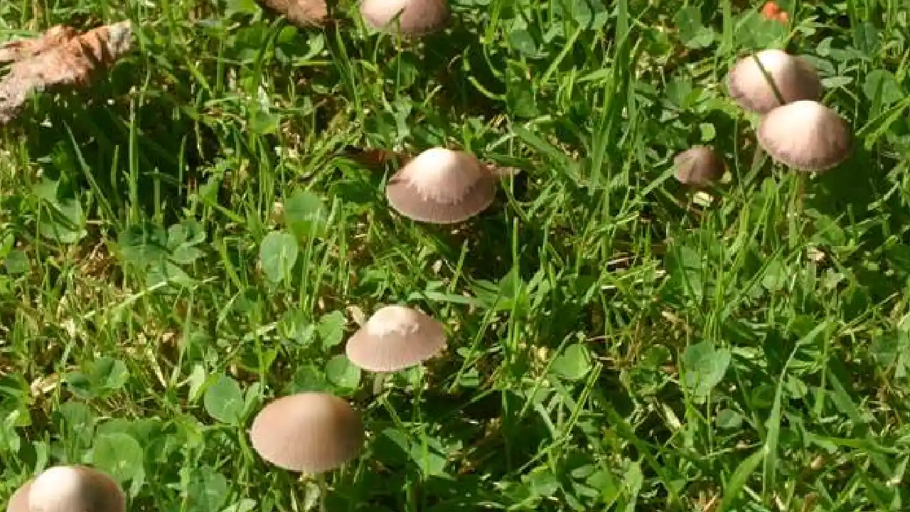
Pholiotina rugosa — The Overlooked Amatoxin Bearer
Pholiotina rugosa — until recently widely known as Conocybe filaris — deserves far more attention than it gets in North American field guides. This is a small, fragile, cone-capped mushroom: pileus typically 1–3 cm, tawny-cinnamon, with a thin stipe bearing a delicate, often evanescent annulus. It grows in lawns, garden beds, wood chips, and disturbed ground across North America.
It contains amatoxins at concentrations comparable to Galerina marginata. A small child eating several fruitings from a suburban lawn has a plausible lethal exposure. The NAMA Toxicology Committee has recorded pediatric cases involving this species — precisely because it grows where children play.
The confusion risk: it resembles dozens of innocuous small brown lawn species — Tubaria furfuracea, Agrocybe praecox, young Marasmius oreades. It's the sort of mushroom that gets ignored because it looks like "just another LBM." That's exactly the attitude that produces emergency room visits.
Spore print is clay-brown to rusty-cinnamon. Stipe is pale with a ring. Pileus is conical to bell-shaped. Find it in your lawn with children or dogs present? Remove it, bag it, and contact your regional Poison Control center if any ingestion is suspected.
Inocybe Species — Muscarine in the Lawn
Inocybe is a massive, taxonomically challenging genus — over 700 described species globally — and a substantial portion are muscarine-positive. Inocybe rimosa (split fibrecap) and Inocybe geophylla (white fibrecap) are among the most common poisoning agents in Europe and both occur across North America.
Physical description of I. rimosa:
- Pileus: 2–6 cm, conical to broadly umbonate, straw-yellow to ochre-brown, with characteristic radial fiber-splits from cap margin toward center — the "rimosa" character
- Stipe: 3–7 cm, pale buff, sometimes with a cortinate zone but no true annulus
- Lamellae: Pallid to clay-brown, fairly crowded, adnate to free
- Spore print: Dull clay to tobacco-brown
- Habitat: Mycorrhizal with both conifers and hardwoods; grasslands, roadsides, gardens; common in urban parks
- Odor: Frequently spermatic or earthy — a genuinely useful character in Inocybe
Muscarine toxidrome onset is rapid — 15 to 30 minutes post-ingestion — and follows the SLUDGE pattern: Salivation, Lacrimation, Urination, Defecation, GI distress, Emesis. Atropine is the antidote, and this is a hospital situation. Rarely fatal in healthy adults if caught quickly. But it's miserable, it mimics cardiac or severe allergic events if the physician doesn't know the exposure history, and children face proportionally greater risk.
The critical lookalike risk: Marasmius oreades grows in the same lawn and grassland habitat with a broadly similar small brown appearance. The separations are firm once you know them — M. oreades has white spores, a tough and pliant stipe, widely-spaced cream-white gills with free attachment, and lacks the fibrous cap texture of Inocybe. Spore print alone breaks the confusion. But novices regularly conflate them, and it's a problem NAMA documents repeatedly across its regional chapters.
Hebeloma crustuliniforme and Paxillus involutus — Underestimated Toxics
These two deserve firm treatment because they're common and frequently minimized in older field literature.
Hebeloma crustuliniforme — poison pie — is a medium-sized LBM (3–8 cm pileus) with a pale buff to honey-brown cap, often with a slight viscid surface in wet weather. Its most useful field character: an unpleasant radish odor, genuinely distinctive, use it. It also shows characteristic droplets of moisture along the gill edge (guttation) in humid conditions. Mycorrhizal with a wide range of trees, common in parks and suburban plantings, and mildly to moderately toxic, causing GI distress. Not a killer. Not acceptable as food.
Paxillus involutus — brown roll-rim — was consumed as a food species in Eastern Europe for generations, and for most of that time the deaths it caused weren't attributed to it. We now understand the mechanism: immune-mediated hemolytic anemia, cumulative in nature. Each ingestion further sensitizes the immune system, and eventually the response to circulating antigens triggers catastrophic red blood cell destruction. People ate it dozens of times before a lethal meal. The German mycologist Julius Schäffer died of it in 1944 — the case that finally clarified the mechanism.
Identifying features: strongly inrolled cap margin in young specimens, brown gills that stain brown-black when bruised or pressed, decurrent gill attachment, rusty-brown spore print, fairly robust at 5–12 cm. Common in birch and conifer woodland across the Northern Hemisphere.
Don't eat it.
Why People Hunt LBMs Anyway — The Edible and Sought Species
Given everything above, why does anyone touch small brown mushrooms? Because some of the most prized or sought species in foraging culture happen to fall in this morphological group — and the people hunting them are often working with higher stakes and more motivation than a casual weekend walk.
Psilocybe semilanceata and P. cubensis
Psilocybe semilanceata — the liberty cap — is one of the most widely distributed psychoactive species in the world. It's a grassland and meadow species, common across the British Isles, Northern Europe, the Pacific Northwest, and parts of the American Northeast. The pileus is distinctively conical to sharply umbonate, rarely exceeding 1.5 cm in diameter, with a characteristic nipple at the apex. The surface is viscid in wet weather, sometimes showing a separable gelatinous pellicle. Spore print: dark purple-brown to near-black. Stipe long and slender, often wavy.
Psilocybe cubensis is primarily tropical to subtropical — Gulf Coast states, Mexico, Central and South America — and is more commonly encountered in cultivation than in wild foraging in North America. In the wild it's a dung-associated species, characteristic of cattle pastures. Both contain psilocybin and psilocin, Schedule I controlled substances in most US jurisdictions.
I've published on the therapeutic potential of psilocybin. I'm not here to adjudicate drug policy. But I'll state clearly: hunting these species creates serious identification risk. People pursuing Psilocybe are often using inadequate field guides in habitats co-inhabited by Galerina marginata, making decisions based on bluing reactions alone.
Bluing is not a reliable Psilocybe indicator under all conditions. Galerina does not blue — but a wet Psilocybe specimen may show weak bluing, or a handled specimen may have already oxidized. The separator is the spore print: purple-brown to black for Psilocybe, rust-brown for Galerina. There is no shortcut. People have died pursuing Psilocybe and ingesting Galerina instead. That's a documented, recurring pattern.
Get the spore print. Every single time.
Kuehneromyces mutabilis — Europe's Fatal Confusion
Kuehneromyces mutabilis — the sheathed woodtuft — is a legitimate, well-regarded edible in continental European cuisine. It grows in dense clusters on dead hardwood logs and stumps, particularly beech and oak. Pileus honey-brown to tawny, strongly hygrophanous with a two-toned appearance — pale at center, darker at margin when drying. Stipe with a distinct annulus and a scaly, dark-brown lower portion below the ring. That scaling is its most reliable field character.
The problem: Galerina marginata grows in almost identical habitat, in almost identical clusters, at the same season, with almost identical pileus coloration. The stipe scaling distinguishes them — present in K. mutabilis, absent in Galerina — but both species produce rusty-cinnamon spores, removing that separator. Microscopy — specifically the presence of chrysocystidia in K. mutabilis versus different cystidia morphology in Galerina — is often needed for certainty.
European poisonings involving this pair are well-documented. With K. mutabilis naturalized in parts of the Pacific Northwest, American foragers should treat any wood-clustered honey-brown LBM as Galerina until proven otherwise by microscopic examination.
Marasmius oreades, Armillaria mellea, and Flammulina velutipes
These three represent the genuinely safe end of the LBM-adjacent edible spectrum — with qualifications.
Marasmius oreades — fairy ring mushroom — is one of the finest edible species in temperate grassland mycology. It grows in characteristic arcs and rings in lawns and meadows, following the line of underground mycelium. Pileus pale tan to buff, 2–5 cm, with a broad central umbo. The most useful character: the stipe is tough, wiry, and pliant — it bends without breaking, unlike most similar-looking species. Gills are cream-white, widely spaced (not crowded), free to nearly free. White spore print.
The lookalike danger is Inocybe species sharing the same lawn habitat. A firm spore print on white paper resolves it: white for M. oreades, clay-brown for Inocybe. Don't skip it.
Armillaria mellea — honey mushroom — is a large-scale cluster former on living and dead wood, causing white rot, and genuinely edible when properly cooked (raw or undercooked consumption causes GI distress in most people). Pileus honey-yellow to tawny-brown with scattered dark scales, 3–12 cm. Stipe tough and fibrous, with a prominent white to pale yellow annulus. White to cream spore print. Black rhizomorphs under bark at the stipe base are often the giveaway in the field.
Flammulina velutipes — velvet shank, or cultivated enoki — is a winter and early spring species distinguished by timing and by its velvety, dark-brown lower stipe. Pileus orange-brown to tawny, viscid. White spore print. It clusters on dead hardwood stumps, often fruiting through snow. Timing and habitat make serious misidentification with toxic LBMs unlikely — but always confirm the spore print.
The Ambiguous Middle — True LBM Grind Species
Here's where I'll be honest with you: these are the species that don't make headlines, don't appear on poisoning registries, and don't have dramatic common names. They're also the species that comprise the majority of small brown mushrooms you'll encounter on any given day in the field. Learning to recognize these genera — even without species-level ID — is a functional skill for any serious forager, because it tells you what you're not looking at.
Pholiota species grow on wood, often in clusters, with distinctively scaly pileus surfaces that may wash off in rain. Spore print rusty to dark brown. Most are bitter or cause GI irritation. Pholiota squarrosa is a common wood-clustered species with prominent dry, recurved scales that novices confuse with Armillaria. Chrysocystidia are present in most species under KOH — a useful microscopic marker that separates them from Galerina.
Hypholoma fasciculare — sulfur tuft — causes enough poisonings to warrant attention. Dense clusters on dead wood, pileus sulfur-yellow to tawny at center with greenish-yellow tones in the flesh. Gills turn olive-green with age. Spore print dark purple-brown. Tastes distinctly bitter. H. capnoides grows in identical habitat and cluster form but has gills that stay grayish-lilac rather than greenish, with no bitter taste — it's edible and reasonably good. Gill color and taste (spit, don't swallow) separate them, but get the spore print regardless.
Tubaria furfuracea — dingy twiglet — is one of those ubiquitous background species: tiny (1–3 cm), fluted or striate at the pileus margin, orange-brown, on twigs and woody debris. It won't kill you, but it's the species that makes beginners realize they're taxonomically in over their heads. No edibility value, but worth learning as a recognition anchor.
Crepidotus species are stemless or nearly so, fan-shaped or kidney-shaped, growing laterally from dead wood. The absence of a proper stipe is usually diagnostic immediately. Spore prints brown. No edibility value, no significant toxicity.
Naucoria species are alder-associated in streamside habitats, particularly the Pacific Northwest and Northern Europe. Pileus brown to ochre, often with a mealy odor. Spore print rust-brown. Taxonomically difficult — largely separated by microscopy. Toxicity unknown for most species, which is itself a sufficient reason not to eat them.
Phaeocollybia species are distinctive for the long, tapering, cartilaginous stipe that continues deep into the substrate as a rooting pseudorhiza. Pacific Northwest conifers, particularly Douglas fir associations. Some species are quite large for LBMs. No established edibility or toxicity for most; taxonomic difficulty is severe without microscopy and DNA.
Gymnopus dryophilus — russet toughshank — is a well-behaved saprobe on leaf litter, common in deciduous woodland from spring through autumn. Pileus russet-brown to pale buff, smooth. Stipe tough and cartilaginous. White spore print — which immediately distinguishes it from most dangerous LBM genera. Worth knowing as a white-spored small brown species in leaf litter.
Simocybe and Xeromphalina campanella round out the group. X. campanella is distinctive for its orange-brown pileus with decurrent gills and tendency to grow on mossy conifer wood in large troops. White to pale yellow spore print. Not dangerous, not edible. A useful reference point for habitat and size.
The practical lesson from this entire category: take a spore print before you key anything further. That single action narrows the field in ways that no amount of cap-color comparison can match.
Field Identification — What to Actually Look For
I've taught university-level mycology. I've supervised hundreds of students learning to identify mushrooms. The single biggest gap I see — in students, in foragers, in online ID communities — is the failure to work systematically through characters in the right order. People photograph the cap, post it to iNaturalist or a Facebook group, and expect an answer. That's not identification. That's a guess with extra steps.
Here's the systematic approach for LBMs, ordered by reliability.
Pileus — Shape, Color, and Hygrophanous Behavior
Start with the pileus — not as a color judgment, but as a structural assessment.
Shape progression: Is it conical and sharply peaked like Psilocybe semilanceata? Convex to broadly umbonate like many Galerina? Plane with a broad central umbo like many Inocybe? Bell-shaped expanding to plane like Hebeloma? The shape at maturity matters, but so does understanding the developmental stage of the specimen you're holding.
Surface texture: Smooth and viscid in wet weather? Dry and fibrous? Distinctively scaly like Pholiota or young Armillaria? Radially cracked like Inocybe rimosa? These characters are more durable than color — they persist through weather changes.
Hygrophanous behavior: Note whether the cap shows a two-toned appearance — darker at the margin where moisture is retained, paler toward the center where it's drying. This is nearly universal in Galerina, Kuehneromyces, Tubaria, and many Cortinarius. When you see it, you're in a group where color-based identification is particularly unreliable.
Size and margin: Under 3 cm puts you in micro-LBM territory — Tubaria, Pholiotina, small Galerina. Striate margin — radiating translucent lines from the edge — indicates thin flesh and close gills, useful in separating from species with opaque margins.
Stipe, Annulus, and Cortina — The Safety-Critical Structures
The stipe is where safety-relevant characters cluster in this group. Examine it with a 10× hand lens. Don't skip this step.
Annulus: Present or absent? If present — membranous and skirt-like (higher concern for Galerina, Pholiotina)? Or merely fibrous remnant zones? The annulus in Galerina marginata is delicate and frequently deteriorated in mature specimens — you may find only fibrous striations where a ring once was. Absence of a ring in an older specimen is not evidence that the species lacks one.
Cortina: The cobwebby, fibrous partial veil unique to Cortinarius. In young specimens it's clearly visible as silky threads spanning from stipe to pileus margin. In older specimens it collapses to rusty-orange fiber zones on the upper stipe, stained by deposited spores. A brown-spored LBM with rusty-orange webby remnants on the upper stipe: you're in Cortinarius, and you do not eat it.
Stipe surface texture: Smooth and pale throughout? Fibrous and longitudinally striate? Scaly in the lower half — a positive feature for Kuehneromyces mutabilis? Velvety and dark brown at the base like Flammulina velutipes? Rooting deeply into the substrate like Phaeocollybia?
Stipe base: Any abrupt bulb? Any cup (volva)? An abrupt marginate bulb moves you immediately toward Amanita territory, away from LBMs — but it's worth noting that boundary explicitly so beginners don't mistake a volva-less Galerina for a safe find.
Gill Attachment and Color Progression
Gill attachment describes how the lamellae connect to the stipe:
- Free: Gills don't touch the stipe — typical of Amanita and some Marasmius; not typical LBM
- Adnexed: Narrowly attached — common in Galerina, Psilocybe
- Adnate: Broadly attached — common across many LBM genera
- Decurrent: Gills run down the stipe — Flammulina, some Gymnopus, Xeromphalina
Gill color across age: Young Galerina gills are pallid to cream; they darken to rusty-cinnamon as spores mature. Hypholoma fasciculare gills shift from pale yellow to distinctively olive-green — a reliable aging character. Inocybe gills begin pallid and become clay-brown. Photograph gills of both young and mature specimens when possible.
Gill spacing: Crowded versus widely spaced is a genuine, durable character. Marasmius oreades has widely spaced, broad, cream gills — distinctive once you've seen it once.
Spore Print — The Single Most Important Step
Do this for every unidentified LBM. No exceptions, no shortcuts.
Remove the pileus, place it gill-side down on a piece of white paper — better still, a sheet divided between black and white sections, since pale prints show on dark and dark prints show on white. Cover with a glass or bowl to prevent airflow. Leave undisturbed for at least 30 minutes; an hour is better.
| Spore Print Color | Primary Genera |
|---|---|
| Rust-brown / cinnamon | Galerina, Cortinarius, Pholiota, Tubaria, Naucoria |
| Dark purple-brown to black | Psilocybe, Hypholoma, Stropharia, Panaeolus |
| Clay / ochre-brown | Pholiotina, Inocybe (some) |
| White to cream | Marasmius, Gymnopus, Flammulina, some Crepidotus |
| Pink to salmon | Entoloma spp. — separate genus, also dangerous |
A rust-brown to cinnamon spore print on a wood-growing small brown mushroom means Galerina until proven otherwise by microscopic examination. That's the operative rule, and nothing in this guide supersedes it.
Chemical Spot Tests
Chemical reagents add another identification layer, particularly useful when microscopy isn't immediately available.
KOH (10% solution): Applied to pileus flesh or surface. Produces color reactions that vary by genus. In some Cortinarius groupings, KOH produces yellow to olive changes on the pileus surface. On Hebeloma, KOH applied to gill surface may produce distinctive reactions. These reactions narrow the genus — they don't determine the species.
Melzer's reagent: Tests for amyloid reaction. Spores or tissue staining blue-black in Melzer's are amyloid; reddish-brown is dextrinoid; no reaction is inamyloid. Galerina spores show characteristic ornamentation and may show dextrinoid reactions. Primarily useful at the microscopic level.
Iron salts (FeSO₄): Flesh color reaction used as a discriminating character in some Cortinarius subgenera. Green staining on Cortinarius flesh with iron salts is reported in certain species complexes — genus-level narrowing, not species determination.
Bluing reaction: Bruise the stipe base or cut through it. Blue-green staining developing within minutes indicates psilocybin content — a positive indicator for Psilocybe and a few related genera. Critically: the absence of bluing does not rule out Galerina. These two species operate in completely different chemical frameworks. Spore print remains necessary regardless of bluing result.
When You Need a Microscope
For any LBM where you want a definitive identification — and especially before any edibility decision — microscopy is not optional. It's the standard.
The microscopic characters that matter in this group:
- Cheilocystidia (gill edge cystidia): Shape, size, and wall structure are often genus- and species-diagnostic. Galerina cystidia differ from Kuehneromyces in ways clearly visible at 400× with appropriate preparation.
- Pleurocystidia (gill face cystidia): Present or absent; shape varies significantly across genera.
- Chrysocystidia: Thick-walled, golden-refractive cystidia in KOH — present in Hypholoma, Stropharia, Pholiota; absent in Galerina. This character alone separates critical lookalike groups.
- Spore ornamentation: Smooth, verrucose (warty), striate, nodulose — visible at 1000× in oil immersion. Cortinarius spores are characteristically verrucose.
- Clamp connections: Presence or absence at hyphal septa — useful generic character across the board.
For serious or ambiguous specimens, ITS DNA barcoding is now the decisive tool. Submit to regional herbaria, university mycology departments, or commercial barcoding services with appropriate documentation. MushroomExpert.com by Michael Kuo and Mushroom Observer both facilitate community-assisted identification with photographic documentation — but neither replaces a spore print, and neither replaces a certified mycologist's opinion before consumption.
If you've consumed an unknown LBM and have any doubt at all: call Poison Control at 1-800-222-1222. That's 24/7, 365 days a year, staffed by toxicologists who know mushroom poisoning cold. Don't wait for symptoms. With amatoxin-producing species, symptoms arrive far too late.
Sections 6 through 9 — habitat profiles, full toxin clinical detail, emergency protocol, and reference resources — follow in the next installment.
Habitat — Where LBMs Actually Grow
Wood Chips and Urban Mulch — The Highest Risk Zone
I want to be direct about something most field guides bury in footnotes: the most dangerous LBM habitat in North America is not a remote wilderness forest. It's the wood chip mulch bed outside your apartment complex, in the median of your city park boulevard, around the ornamental shrubs at a hospital entrance.
Galerina marginata — and to a lesser extent Pholiotina rugosa — thrives in commercially produced wood chip mulch. The chips are almost always softwood or mixed wood, exactly the substrate these species evolved to decompose. Urban mulch beds retain moisture, warm quickly in spring, and extend fruiting seasons well beyond what you'd see in natural forest. I've pulled Galerina from wood chip beds in October rain in Seattle, in March thaw in Portland, and in humid November in cities across the South.
The urban risk compounds because of accessibility. Children play near ornamental plantings. Dogs root through mulch. Foragers who feel confident in wild woodland let their guard down in city parks. And Galerina fruitings in urban mulch are often dense — thirty or forty fruiting bodies in two square meters, meaning a single accidental ingestion can represent a significant amatoxin load.
Psilocybe cyanescens and P. azurescens on the West Coast also fruit prolifically in urban wood chip beds — which is precisely why this habitat matters so much. People hunting Psilocybe in city park mulch are foraging in the exact same substrate where Galerina is fruiting. I've surveyed both species in the same 10-square-meter plot. The consequence of a field mix-up in that context is liver failure.
If you're introducing anyone — children, newcomers, enthusiastic beginners — to mushroom foraging, start with this: a wood chip bed is not a safe foraging environment. It looks benign. It isn't.
Pacific Northwest Conifers
The Pacific Northwest — from Northern California through Oregon and Washington into British Columbia — is arguably the richest fungal territory in the Northern Hemisphere. Douglas fir (Pseudotsuga menziesii), Sitka spruce (Picea sitchensis), western hemlock (Tsuga heterophylla), and western red cedar (Thuja plicata) create a dense, humid canopy under which the fungal biomass is extraordinary.
For LBMs specifically, this region presents a demanding and layered identification environment:
- Galerina marginata fruits from dead conifer logs and stumps throughout a season that runs September through February in mild coastal years
- Phaeocollybia species — rooting-stipe brown mushrooms largely unique to this region — associate primarily with Douglas fir and hemlock; several are taxonomically undescribed and require DNA for confident placement
- Cortinarius is the dominant genus by biomass in old-growth and second-growth stands; the Pacific Northwest hosts hundreds of species, many rust-brown and medium-sized, resistant to separation without microscopy
- Psilocybe cyanescens and P. allenii fruit in disturbed woodland, riparian zones, and wood chip beds throughout the lowland coastal strip — directly overlapping in habitat and season with Galerina
That seasonal overlap is the practical danger. Peak Cortinarius fruiting in the Cascades runs September through November. That's also peak P. cyanescens season in lowland western Oregon and Washington. And peak Galerina season in coastal forest. Three groups, radically different risk profiles, fruiting simultaneously in habitats a short drive apart.
I've collected in the Olympics, the Cascades, and the coastal rainforests of Oregon for decades. My rule in Pacific Northwest conifer forest: any rust-brown small mushroom on or near wood gets a spore print before any other assessment. The diversity here is breathtaking, and its LBM component is accordingly unforgiving.
Appalachian Hardwoods
The Appalachian corridor — running from Georgia and the Carolinas through Virginia, Tennessee, West Virginia, Pennsylvania, and into New England — presents a different fungal character entirely. The dominant overstory is mixed hardwood: white oak (Quercus alba), red oak (Q. rubra), American beech (Fagus grandifolia), sugar maple (Acer saccharum), and at lower elevations, tulip poplar (Liriodendron tulipifera).
Key LBM dynamics in this region:
- Armillaria mellea and its close relatives are aggressively common on dead hardwood, particularly oak and beech; I've pulled clusters from the same white oak stumps four autumns running in western North Carolina
- Galerina marginata is present on dead wood but less dominant than in Pacific Northwest conifers — still a genuine risk, never dismissible
- Hebeloma crustuliniforme is widespread in autumn, mycorrhizal with both oak and beech, appearing in enormous numbers after soaking rains in September and October
- Inocybe rimosa and related fibrecap species are particularly well-represented in oak-associated grassland edges and disturbed soils through the southern Appalachians
- Agrocybe praecox and A. pediades — spring and early summer species — are common in disturbed ground and wood chip areas across the region
The Appalachian season runs later than most foragers expect. I've found productive Armillaria flushes in Tennessee in late November, with Galerina active alongside them on adjacent logs. The beech-maple forest floors of western North Carolina and the Blue Ridge produce extraordinary LBM diversity that remains substantially under-surveyed. The Virginia Mycological Society and regional forays organized through NAMA affiliates are your best entry point for local knowledge in this corridor.
Grasslands, Lawns, and Meadows
Open habitat — lawns, meadows, golf courses, cattle pastures, roadsides — is where the public most routinely encounters small brown mushrooms, and where the identification errors that reach emergency rooms most often originate. There's a false safety in open ground. People assume danger lives in dark forest. It doesn't.
Key species in this habitat:
- Marasmius oreades — the primary edible target, producing its characteristic arcs and rings, but fruiting in the same grassland at the same season as Inocybe rimosa and I. geophylla
- Psilocybe semilanceata — in the Pacific Northwest and across the British Isles, specifically in ungrazed or lightly grazed permanent pasture, often associated with Agrostis grass species and dung-enriched soils
- Pholiotina rugosa — suburban and urban lawns, garden beds, enriched soils; the amatoxin-bearing lawn species that most people have never heard of
- Panaeolus species — dung-associated, common in cattle and horse pastures; not typically dangerous, but identification within the genus requires care
- Conocybe species — fragile, conical, and common in lawns; mostly innocuous, though the Pholiotina/Conocybe taxonomic boundary is genuinely messy and warrants caution
The fairy ring is a useful habitat feature but a misleading identification cue. Marasmius oreades produces classic rings where the mycelium exhausts soil nutrients, creating dark-green grass at the perimeter and sometimes bare ground at the center. But Inocybe species, Clitocybe species, and Lepista species all produce fairy rings independently. The ring shape alone tells you nothing useful about species.
In cattle pasture specifically: dung-associated Psilocybe and Panaeolus occupy overlapping habitat, the Inocybe risk from adjacent woodland edges is real, and the complacency that flat, open terrain breeds in foragers is itself a hazard. Same rule applies as everywhere else: spore print before any further conclusions.
Dead Logs, Stumps, and Streambanks
Dead wood is the most species-rich LBM habitat, and for practical purposes it's where the most dangerous misidentifications cluster. The saprotrophic LBMs decomposing dead wood — Galerina, Hypholoma, Kuehneromyces, Pholiota, Crepidotus, Flammulina — share this substrate with edible species like Armillaria and Pleurotus. Reward and risk grow on the same log. Literally.
Key patterns:
- Conifer logs in the Pacific Northwest and boreal forest: Galerina marginata and Hypholoma capnoides are the most common fruiters. H. capnoides is edible and good; Galerina kills. They share substrate, season, and a broadly similar small brown habitus. Spore print separates them — dark purple-brown for H. capnoides, rust-cinnamon for Galerina.
- Hardwood stumps in eastern North America: Armillaria complex species, Hypholoma fasciculare, and — increasingly significant as it naturalizes in the Northwest — Kuehneromyces mutabilis all cluster on hardwood. Flammulina velutipes takes over the same stumps in winter when competitors have stopped fruiting.
- Alder streambanks in the Pacific Northwest: Naucoria species associate specifically with alder (Alnus spp.) roots in riparian zones. They're a genuinely regional LBM challenge — taxonomically understudied, not safely edible, and common enough that streamside foragers encounter them regularly.
- Apparently terrestrial fruitings near wood: Galerina frequently fruits from what looks like bare soil when the actual substrate is a buried root or log fragment inches below the surface. Don't let the absence of visible wood reassure you.
Phaeocollybia species deserve a specific note for streambank habitats. They fruit in the root zones of conifers adjacent to waterways across the Pacific Northwest, and the rooting stipe is diagnostic — pull gently upward and you'll feel the pseudorhiza anchoring deep into the substrate. Striking organisms. No established edibility, no established toxicity, no reason to eat them.
The principle for all dead wood foraging: cluster growth on wood, plus brown pileus, equals immediate spore print before anything else. Armillaria and Galerina can fruit centimeters apart on the same log. I've seen it more than once. Don't assume cluster means safety.
Toxin Profiles and What Happens If You're Wrong
The species introductions in Section 2 established which compounds are involved. Here I want to go deeper — into mechanism, clinical reality, and what treatment actually looks like — because this is the information that matters when something has already gone wrong.
Amatoxins — Mechanism, Organ Failure, and Treatment
Amatoxins are bicyclic octapeptides. The primary toxic compound in Galerina marginata and Pholiotina rugosa is α-amanitin, with β-amanitin and γ-amanitin present in supporting roles. Their mechanism is precise and lethal: they bind to and inhibit RNA polymerase II, the enzyme responsible for transcribing DNA into messenger RNA. Without functional mRNA transcription, protein synthesis collapses. The cells most vulnerable are those with the highest metabolic demand — hepatocytes first, then renal tubular cells and GI epithelium.
Critically: amatoxins are not inactivated by cooking, drying, or freezing. They're water-soluble, which means the broth from a pot of cooked Galerina is as toxic as the fruiting body itself. There is no preparation method that renders these mushrooms safe.
Lethal dose context: The estimated lethal dose of α-amanitin in humans is approximately 0.1 mg per kilogram of body weight. A single fresh cap of Galerina marginata can contain 0.01 to 0.3 mg of total amatoxin depending on specimen size and growing conditions. A 15-kilogram child could receive a lethal dose from one small fruiting body. This is why the urban wood chip bed exposure I described in Section 6 is a pediatric risk, not a theoretical one.
The false recovery phase — apparent clinical improvement around days 2 to 3 — is the feature that historically caused the most deaths before the mechanism was understood. Patients felt better, were discharged, and presented in fulminant liver failure two days later. Modern toxicology centers know to maintain observation and serial hepatic function monitoring through this window, regardless of apparent improvement.
Current treatment protocol:
| Intervention | Timing | Mechanism |
|---|---|---|
| Activated charcoal | Within 1–2 hours of ingestion | Interrupts enterohepatic amatoxin recirculation |
| IV fluid resuscitation | Immediate | Renal protection, hemodynamic support |
| Silibinin IV (Legalon SIL) | As early as possible | Competes with OATP-mediated hepatocyte uptake — the closest thing to a specific antidote |
| N-acetylcysteine | Early to mid-course | Glutathione replenishment, hepatocyte oxidative protection |
| Liver transplant evaluation | Day 3–4 if labs trending badly | PT/INR, bilirubin, encephalopathy are the triggers; don't wait for day 5 |
Silibinin IV — derived from Silybum marianum, milk thistle — is available in the US through compassionate use protocol. Early administration substantially improves outcomes. If your treating physician isn't familiar with it, the NAMA Toxicology Committee and Poison Control can facilitate access information.
What to bring to the ER: A sample of the mushroom in a paper bag — not plastic, which accelerates decomposition. A spore print if you have one. Photographs. Whatever the patient didn't eat from the same collection. This material can reach a consulting mycologist within hours through the NAMA Toxicology network.
Orellanine — The 21-Day Delay That Kills Kidneys
Orellanine — the nephrotoxic compound in Cortinarius rubellus, C. orellanus, and related webcap species — is in some ways the most insidious mushroom toxin known. It's a bipyridyl compound that operates as a photosensitizer: it absorbs UV radiation and generates reactive oxygen species in the presence of light. In renal tubular cells, this produces progressive oxidative destruction of the tubular epithelium.
The latency of two to twenty-one days is not a rough estimate. It's documented across European case series — particularly from Poland, Germany, and the Czech Republic, where C. orellanus is a common woodland species. Cases on record include patients who presented to nephrology clinics weeks after a meal, with no initial connection between cause and effect, until a careful dietary history surfaced the Cortinarius exposure.
Clinical presentation: Intense thirst, polyuria as damaged tubules lose their concentrating ability, flank pain, nausea, progressing to oliguria as renal failure establishes. Creatinine and blood urea nitrogen climb. In severe documented cases, dialysis was required; renal transplant has been performed.
There is no antidote. No orellanine-specific therapy exists. Everything is supportive. And the kidney damage is often irreversible — some patients retain residual function after the acute phase; others require long-term dialysis.
What makes orellanine uniquely dangerous in the foraging context: Cortinarius species are common, visually unremarkable, and don't cause acute illness. There's no warning system. A forager who eats Cortinarius rubellus in October and feels perfectly well has no reason, without specific knowledge of this toxin, to associate a November kidney crisis with a meal weeks earlier.
If you've eaten any Cortinarius species and cannot confirm identification by microscopy or DNA barcoding: call Poison Control at 1-800-222-1222 and name the genus and the date of ingestion. Even if you feel completely fine. Especially if you feel fine.
Muscarine — Fast Onset, Manageable If Caught Early
Muscarine — concentrated in Inocybe species and to a lesser extent in some Clitocybe — produces a rapid, unambiguous clinical syndrome that is simultaneously this toxin's most alarming feature and its most tractable one. Unlike amatoxins and orellanine, muscarine poisoning announces itself quickly. There's time to intervene effectively.
Muscarine is a quaternary ammonium compound that mimics acetylcholine at muscarinic receptors without being metabolized by acetylcholinesterase. The result is sustained cholinergic stimulation across all parasympathetic end organs simultaneously.
| Symptom | Mechanism |
|---|---|
| Salivation, lacrimation | Exocrine gland hyperstimulation |
| Urination, defecation | Smooth muscle hyperstimulation |
| GI cramping, emesis | Intestinal and gastric hypermotility |
| Bradycardia | Cardiac vagal stimulation |
| Bronchospasm | Pulmonary smooth muscle constriction |
| Miosis | Pupillary sphincter contraction |
Onset is 15 to 30 minutes post-ingestion — fast enough that most patients remember precisely what they ate and when. That history is what makes treatment immediate and effective.
Treatment: IV atropine — a muscarinic antagonist — is the definitive intervention and highly effective. Dose titration targets resolution of bronchospasm and excess secretions; the goal isn't full atropinization, which causes its own toxidrome. Recovery is generally complete in healthy adults within 12 to 24 hours of appropriate treatment.
The risk population that changes the calculus: Children, elderly patients, and anyone with underlying cardiac or pulmonary disease face disproportionate risk from even moderate muscarine exposure. A healthy adult presenting to the ER within two hours of eating a few Inocybe caps is going to be deeply uncomfortable but almost certainly recovers fully. A 70-year-old with COPD in the same situation is a genuine emergency with airway implications.
The practical information for the ER: muscarine, atropine, 2–4 mg IV, titrate to secretions. Name the genus — Inocybe — and name the toxin. If the treating physician knows it, treatment starts immediately. If they don't, that information shortens the path considerably.
If You've Eaten Something — Emergency Protocol
I've consulted on mushroom poisoning cases for emergency rooms across multiple states over four decades. The single most important variable in patient outcome isn't the species, isn't the dose, and isn't the patient's age. It's how quickly the right physician gets the right information.
Here's what to do, in order. Don't improvise. Don't wait.
Call Poison Control immediately: 1-800-222-1222.
This call is free, available 24 hours a day, 365 days a year, and staffed by toxicologists who handle mushroom cases. They'll ask what was eaten, how much, when, and what symptoms if any are present. They'll help determine whether you watch and wait or go to the ER right now. For any ingestion involving a small brown mushroom you cannot positively identify — go to the ER. The watching and waiting option doesn't apply here.
Collect your evidence before you leave.
- Save any uneaten portion of the mushroom in a paper bag — not plastic, which accelerates decomposition and makes visual and chemical ID harder
- Photograph the collection site if you can return quickly or have photos from earlier
- Save any vomit if vomiting has already occurred — it may contain spores or tissue fragments sufficient for identification
- Bring the spore print if you made one
- Document the time of consumption, approximate quantity, and preparation method
Go to the emergency room — even without symptoms.
This is non-negotiable for any unknown small brown mushroom ingestion. The latency periods for amatoxins (6–24 hours) and orellanine (days to weeks) mean that feeling fine right now is not reassurance. At the ER, ask specifically for a toxicology consult. Tell them:
- The genus name if you know it — or "unknown small brown mushroom, rust-brown spore print" if you don't
- The exact time of ingestion
- That your concern is amatoxin-producing species in the Galerina or Pholiotina complex
Liver function tests — ALT, AST, bilirubin, PT/INR — should be drawn on arrival and monitored serially for at least 72 hours in any suspected amatoxin case. Normal initial labs are encouraging but not conclusive. Repeat them.
Involve the NAMA Toxicology Committee.
The North American Mycological Association maintains an emergency toxicology consultation service specifically for mushroom poisoning cases. The committee includes practicing toxicologists and experienced mycologists who can assist with rapid species identification and advise on clinical management. Your ER physician can reach them through the Poison Control network or directly through NAMA. A specimen or clear photographs dramatically accelerate this process — which is why the collection evidence step above is not optional.
What not to do:
- Don't induce vomiting unless Poison Control explicitly instructs you to — aspiration risk is real
- Don't wait for symptoms before calling or going in
- Don't let an online identification convince you the species was safe
- Don't assume cooking neutralized the toxins — amatoxins are heat-stable and water-soluble
- Don't treat a history of eating this before without problems as evidence of safety — Paxillus involutus kills through cumulative immune sensitization; previous safe exposures are not protection against the next one
My mantra holds: there are old mushroom hunters, and there are bold mushroom hunters, but there are no old, bold mushroom hunters. The emergency protocol exists because the biology is unforgiving. Use it.
Tools, References, and Where to Learn More
Identifying small brown mushrooms well requires a layered toolkit. No single resource — not a field guide, not an app, not an online community — is sufficient on its own. Here's what I actually use and recommend, in order of reliability.
Primary Identification Tools
Spore print and hand lens: The non-negotiable starting point. A quality 10× hand lens costs less than a restaurant meal and reveals annulus remnants, cap surface texture, and gill structure invisible to the naked eye. It belongs in your field kit on every outing.
Compound microscope: For serious LBM work, a microscope capable of 400× and 1000× (oil immersion) is the tool that separates genera and resolves species. A basic student-grade scope is adequate to begin — you don't need research-grade optics to examine cheilocystidia or read spore ornamentation. Regional mycological societies run microscopy workshops worth attending.
Chemical reagents: KOH (10% solution), Melzer's reagent, and iron salts are all commercially available or inexpensively prepared. A small field reagent kit extends your toolkit considerably — particularly useful for Cortinarius discrimination with iron salts and Hypholoma versus Galerina separation using chrysocystidia detection in KOH.
ITS DNA barcoding: Now accessible and affordable through university herbarium programs and commercial services. Submit a dried specimen with complete collection data — GPS coordinates, associated tree species, substrate, date — and receive sequence results that can place the specimen to genus or species level. For ambiguous LBMs, this is the definitive tool. It's what I use when macroscopic and microscopic characters don't converge on a clean answer.
Field Guides Worth Owning
David Arora, Mushrooms Demystified (1986): Still the most comprehensive single-volume treatment of North American mushrooms in print. Arora's coverage of Galerina, Inocybe, and Cortinarius is honest about identification difficulty in ways many more recent guides avoid. Required on any serious mycologist's shelf — even if it now needs updating on certain taxonomy.
McKnight and McKnight, A Field Guide to Mushrooms of North America (Peterson Field Guides): More portable than Arora, with solid regional coverage and appropriately conservative edibility notes. A good field companion for the pack.
Gary Lincoff, National Audubon Society Field Guide to North American Mushrooms: Strong photographic coverage, useful for visual comparison alongside written keys. Never use photographs as sole identification evidence — use them as one layer in a multi-character approach.
Breitenbach and Kränzlin, Fungi of Switzerland, Volumes 4 and 5: The most rigorous treatment of agarics in any accessible format. Covers European species but is directly relevant for Cortinarius, Inocybe, Hebeloma, Galerina, and Pholiota — genera with broad Northern Hemisphere distributions. Expensive and hard to source new; worth hunting down through specialist booksellers for serious students.
My own Psilocybin Mushrooms of the World (1996): Relevant specifically for anyone working in the Psilocybe space, with detailed coverage of the Galerina marginata lookalike risk and spore print differentiation. The identification rigor there was deliberate — I knew exactly what the stakes were.
Organizations That Matter
NAMA (North American Mycological Association): The primary national body for amateur and professional mycologists in North America. NAMA maintains the Toxicology Committee, organizes regional forays, publishes McIlvainea, and maintains a national network of experienced mushroom identifiers. Membership gives you access to that network. Find your regional affiliate — most major metropolitan areas have one, and the regional society foray is the single best learning environment available.
Regional mycological societies: The Oregon Mycological Society, Puget Sound Mycological Society, Boston Mycological Club, New York Mycological Society, and dozens of others run regular forays, microscopy workshops, and expert identification sessions. There's no substitute for going into the field with people who've spent twenty years in your specific regional mycobiota. If you're serious about LBMs in your area, join your local society before eating anything you're uncertain about.
Poison Control — 1-800-222-1222: Not a learning resource in the traditional sense, but the most important contact in this entire article. Save that number in your phone right now. The toxicologists on that line know mushroom poisoning. They will not judge you for calling before any symptoms appear. Call early — that's the whole point.
The Right Learning Sequence
If I were starting from zero and wanted to work safely toward competent LBM identification, here's the order I'd follow:
- Join a regional mycological society and attend a full season of organized forays before foraging solo
- Read Arora cover to cover — not as a field guide to consult in the field, but as a course in fungal literacy
- Learn to take and read spore prints reliably before examining any other character on an unknown LBM
- Build firm recognition of the five most dangerous LBMs in your specific region before learning anything about edible species in this group
- Acquire a basic compound microscope and learn to mount gill sections and read cystidia at 400×
- Only after completing steps 1 through 5: begin working toward species-level identification within specific LBM genera you're genuinely interested in
Skipping any step is how people end up in emergency rooms. The biology doesn't care how confident you are. It cares whether the identification was correct.
The fungal kingdom has been doing this for 450 million years. We've been studying it seriously for perhaps 150. A little humility costs nothing. In the case of small brown mushrooms, it can be the thing that keeps you alive.
Sign in to leave a comment and join the discussion.
 Guide
GuideMedicinal mushroom identification means accurately recognizing fungi with proven health-supporting compounds —and distinguishing them from toxic lookalikes....
 Guide
GuideHow Many Mushroom Gummies Should I Eat: A Practical Guide Straight answer: how many mushroom gummies should i eat depends entirely on what's actually in the...
 Guide
GuideWhy Odor and Texture Matter in Mushroom ID? Odor and texture are two of the most reliable tools in mushroom identification — and experienced foragers treat...