Why Odor and Texture Matter in Mushroom ID?

Nathan Cole
Mycologist · Author · Fungi Expert
Updated
Apr 29, 2026
Why Odor and Texture Matter in Mushroom ID?
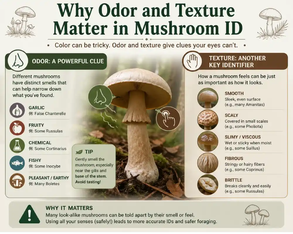
Odor and texture are two of the most reliable tools in mushroom identification — and experienced foragers treat them as mandatory, not optional. When a cap looks identical on two species but one smells like anise and the other like phenol, that single difference can separate an edible mushroom from a toxic one.
Alongside visual features like spore print color and gill structure, organoleptic properties — what you smell, feel, and sometimes taste — form a critical second layer of confirmation. Skipping them is how poisonings happen.
1. The Role of Senses in Mushroom Identification
Most beginner foragers rely almost entirely on what they can see: cap color, gill color, stem shape, habitat. That approach works fine until it doesn't — and when it fails, the consequences can be fatal.
1.1 Why Visual ID Alone Is Risky
Fungi are deceptive. Many species within the same genus look nearly identical under field conditions. Lighting changes cap color. Age warps shape. Rain makes a dry cap look viscid. A photograph in a field guide was taken in different soil, different season, different light.
Consider Amanita phalloides — the Death Cap. It looks unremarkable. Pale, nondescript, easy to confuse with several edible species. Its smell alone won't save you either, because it smells relatively mild. But here's the point: foragers who use only visual cues have no fallback when the visual cues overlap. Every additional sensory data point — smell, texture of the flesh, feel of the cap surface, presence of latex — narrows the ID window.
The multi-characteristic rule is simple:
- One matching feature = interesting
- Three matching features = probable
- Five matching features across visual + organoleptic = confident
1.2 Organoleptic Properties as a Second Layer of Confirmation
Organoleptic refers to anything perceived by the senses — smell, texture, taste, and even sound (does the flesh snap cleanly or tear fibrous?). In mycology, organoleptic testing is a standard part of field identification used in professional keys and academic literature.
The two most practically useful organoleptic properties are:
| Property | What You're Testing | Why It Matters |
|---|---|---|
| Odor | Volatile organic compounds released by the fruiting body | Separates species that look identical visually |
| Texture | Surface feel, flesh structure, gill consistency | Confirms genus-level features, flags dangerous lookalikes |
Together, they function like a fingerprint check after a face match. Visual ID gets you to the right neighborhood. Organoleptic ID gets you to the right house.
2. Understanding Mushroom Odor
Smell is chemistry. When you detect the scent of a mushroom, you're detecting volatile organic compounds (VOCs) — small molecules that evaporate off the mushroom's surface and bind to olfactory receptors. Different species produce different VOC profiles, which is why smell is species-specific enough to function as an ID tool.
2.1 How Volatile Organic Compounds (VOCs) Create Smell
Mushrooms produce VOCs through secondary metabolic pathways — chemical processes that aren't directly tied to growth or reproduction but serve as communication signals, defense mechanisms, and ecological attractants. The VOC profile of a mushroom is partly genetic (species-level) and partly influenced by substrate, age, and environment.
The primary compound responsible for what most people recognize as "mushroom smell" is 1-octen-3-ol, an eight-carbon alcohol also known as mushroom alcohol. It's produced when linoleic acid — a fatty acid in fungal cell membranes — breaks down. Nearly every fleshy mushroom produces it to some degree.
On top of that baseline, individual species layer additional compounds that create distinct scent profiles:
- Benzaldehyde → almond / marzipan scent
- Anisaldehyde → anise / licorice scent
- Geosmin → deep earthy, petrichor-like note
- Phenols → chemical, medicinal, ink-like odor
- Sulfur compounds → garlic, radish, or onion notes
2.2 Common Odor Profiles and What They Indicate
Learning odor profiles is like learning wine notes — it takes repetition, but once the association is locked in, it's reliable across hundreds of specimens.
Anise / Almond (Benzaldehyde + Anisaldehyde) This is a pleasant, sweet smell that immediately points toward specific edible species. Agaricus subrufescens (the almond mushroom), Clitocybe odora (the anise funnel), and Hygrophorus agathosmus all carry this profile. When you smell anise in the field, your attention should snap to these genera.
Mealy / Floury Often described as freshly ground flour or cucumber. Common in Clitocybe, some Entoloma, and notably in Russula and Tricholoma species. The mealy smell comes from compounds called aliphatic aldehydes. Not dangerous on its own, but useful — if a mushroom smells mealy and has white spores and decurrent gills, you're likely in Clitocybe territory.
Fishy / Chemical / Phenolic These are warning smells. A sharp, chemical, or fishy odor often signals compounds that indicate either toxicity or at minimum inedibility. The phenolic smell in Agaricus xanthodermus is one of the most important odor-based warnings in European and North American foraging.
Garlic / Radish / Sulfurous Marasmius scorodonius (garlic parachute) smells so strongly of garlic it was historically used as a seasoning. Radish smell is common in some Russula species and certain Mycena. These sulfur-based VOCs are structurally similar to compounds found in alliums.
Honey / Sweet / Fruity Found in some Armillaria (honey fungus) species and certain Tricholoma. Fruity esters — the same class of compounds that make fruit smell like fruit — appear in scattered species and are usually pleasant but not always edible.
2.3 Key Compound: 1-octen-3-ol (The Classic Mushroom Smell)
1-octen-3-ol deserves its own section because it's the reference point for mushroom odor. It's the compound that makes a mushroom smell like a mushroom — that earthy, slightly damp, fungal baseline you detect even in supermarket varieties.
In the field, high concentrations of 1-octen-3-ol often correlate with mature or slightly past-prime specimens. Young, firm mushrooms may smell subtler. If a mushroom has a very strong, generic "mushroom" smell with no other distinguishing notes, 1-octen-3-ol is likely dominant and you need other features to ID it.
2.4 Dangerous Odors to Know
Phenolic smell in Agaricus xanthodermus — This is the most practically important odor warning for foragers targeting edible Agaricus species. A. xanthodermus (the yellow-staining mushroom) looks nearly identical to Agaricus campestris and Agaricus bisporus relatives. But when you cut or bruise the base of the stem, it releases a sharp, inky, chemical phenolic odor — sometimes described as hospital antiseptic or coal tar.
This smell test is fast, reliable, and saves many foragers from a very unpleasant GI experience every season. The rule: if any Agaricus smells chemical or medicinal at the stem base after cutting, put it down.
Other odor warnings:
- Chlorine / bleach smell → some toxic Tricholoma species
- Rancid / rotten → overripe, bacterial contamination, or certain inedible species
- Musty + sweet together → some Cortinarius species, a genus with dangerously toxic members
3. Odor as a Genus/Species Identifier
At the genus level, odor isn't just a supporting clue — in some cases it's the primary diagnostic feature that keys split on. Here are the most important genus-level odor relationships.
3.1 Agaricus — The Anise vs. Phenolic Split
Agaricus is one of the most foraged genera in the world. It contains excellent edibles (A. campestris, A. bitorquis, A. subrufescens) and one notably toxic species (A. xanthodermus) that causes GI illness in a significant percentage of people who eat it.
The split is almost entirely odor-based:
| Odor at stem base | Species direction | Edibility |
|---|---|---|
| Anise / almond | A. subrufescens, A. silvicola group | Generally edible |
| Neutral / earthy | A. campestris, A. bisporus relatives | Generally edible |
| Chemical / phenolic | A. xanthodermus | Toxic — avoid |
The test: cut the stem base cleanly with a knife and smell immediately. The phenolic smell in xanthodermus is immediate and unmistakable once you've encountered it. It also yellows distinctly when cut — but the smell comes first.
3.2 Inocybe — Sperm-like Odor as a Warning Sign
Inocybe is a large genus of small-to-medium brown mushrooms that are collectively one of the most dangerous groups for beginner foragers. They contain muscarine and other toxins. Almost all should be considered inedible or toxic.
Their most consistent field feature? A distinctive sperm-like or spermatic odor — a sharp, almost chemical-biological smell that most people find instantly unpleasant. Some species smell mealy instead, and a few are nearly odorless, but the spermatic odor in Inocybe is so consistent that experienced mycologists treat it as a genus-level flag.
If you find a small brown mushroom with fibrous cap and that unmistakable smell → do not eat it.
3.3 Clitocybe & Tricholoma — Mealy Smell Pattern
Both genera frequently produce the mealy/floury odor described earlier. In Clitocybe, this combined with crowded, decurrent gills and a funnel-shaped cap narrows the field considerably. In Tricholoma, mealy smell combined with sinuate gills, white to grey coloring, and terrestrial habit in conifer or mixed forest is a strong pattern.
Importantly, Tricholoma equestre (man-on-horseback) — long considered edible — has caused fatalities linked to rhabdomyolysis. Its smell is described as mealy-floury with a slightly sweet note. The lesson: odor helps with ID but does not equal edibility confirmation.
3.4 Cortinarius — Smell as Supporting Evidence
Cortinarius is the largest genus of gilled mushrooms and contains some of the most deadly species in the world — including C. rubellus and C. orellanus, which cause delayed kidney failure through orellanine toxin. The insidious thing about Cortinarius poisoning is that symptoms appear days to weeks after eating.
Odor in Cortinarius is variable — some smell of radish, honey, or have a faint sweetish note. It's not reliable enough to identify or rule out species in this genus. The main organoleptic feature that helps is the cortina — a cobweb-like partial veil connecting the cap edge to the stem in young specimens. It's a texture/structural feature, not a smell. In Cortinarius, always treat smell as supporting data only, never primary.
4. Understanding Mushroom Texture
Texture in mushroom ID operates on three levels: the cap surface, the internal flesh, and the gills. Each level provides different diagnostic information and some features are unique to specific genera.
4.1 Cap Surface Textures and What They Mean
The cap surface (pileus surface) is the first thing you touch in a field examination. Run your finger across it, press lightly, check if it peels, check if it's sticky.
Viscid / Glutinous / Slimy A viscid cap has a sticky or slimy surface, usually due to a gelatinized cuticle layer that absorbs moisture. This is highly genus-relevant:
- Suillus species (bolete relatives in pine forests) are almost always viscid
- Gomphidius and Chroogomphus are typically viscid
- Limacella has a distinctive slimy cap
- Many Cortinarius subgenera (Phlegmacium, Myxacium) have viscid to glutinous caps
A glutinous cap in a gilled mushroom in conifer forest is a strong flag for Cortinarius — remember why that matters.
Dry / Fibrous / Scaly Dry caps are the most common. But the quality of "dry" varies:
- Smooth and dry (Russula, many Agaricus)
- Silky-fibrous (Tricholoma, Inocybe)
- Scaly or squamulose — scales that can be upturned (Pholiota, Lepiota, Amanita species with warty cap remnants)
- Powdery or granular (Amanita exannulate species in the Amanita section)
The presence of warts on the cap in Amanita is actually remnants of the universal veil — a completely different structure than true surface texture. Knowing the difference matters: warts wash off in rain, smooth caps don't always mean no-Amanita.
Hygrophanous This is a crucial and underappreciated texture property. A hygrophanous cap changes color as it dries — typically from darker when wet to paler when dry, often in a water-stain pattern radiating from the center outward.
Species commonly hygrophanous:
- Many Galerina (deadly — amatoxins — often confused with Pholiota or edible small browns)
- Kuehneromyces species
- Some Psathyrella and Hypholoma
If you find a small brown mushroom with a water-stained, two-toned cap, treat it as potentially Galerina until proven otherwise.
4.2 Flesh Texture as an ID Feature
Brittle vs. Fibrous Flesh This is one of the most genus-definitive texture tests in mycology. Take a piece of the cap or stem and try to tear it:
Brittle, crumbly flesh that breaks like chalk → Russula. This genus has a unique tissue structure called sphaerocysts — rounded cells instead of the elongated hyphal cells in most fungi. The result is flesh that snaps, doesn't string, and crumbles when you rub it between your fingers. No other common gilled mushroom genus has this texture.
Fibrous, stringy flesh that tears like chicken or string cheese → most other genera, including Tricholoma, Agaricus, Clitocybe, Cortinarius
If you find a brightly colored gilled mushroom (red, yellow, purple, green) with brittle, non-fibrous flesh → you're almost certainly in Russula. This single texture test is faster and more reliable than color alone in this genus.
Hollow vs. Solid Stipe Cut the stem lengthwise:
- Hollow from top to base → Marasmius, some Mycena, Pluteus, many Psathyrella
- Stuffed / pithy → Russula, Lactarius, older specimens of many species
- Solid and dense → Cantharellus, Craterellus, many Agaricus
A hollow stipe in a larger, fleshy mushroom rules out many edible species and is worth noting.
Cottony / Stuffed Interior Some species have a stipe filled with loose cottony pith — Macrolepiota and Lepiota species often show this. In some Agaricus, the stipe base may have a thick, cottony ring zone. These internal texture features matter in keys.
4.3 Gill Texture
Gills are where some of the most important texture distinctions live.
Waxy Gills (Hygrophorus / Hygrocybe) Waxy gills feel exactly like what they sound like — thick, waxy, like candle wax or wet soap. Rub a gill between your fingers; it should smear slightly rather than crumble. This texture is diagnostic for the family Hygrophoraceae.
Hygrophorus and Hygrocybe (waxcaps) are among the most ecologically important fungi in ancient grasslands and old-growth forests. Their waxy gills combined with bright coloring (scarlet, yellow, orange) make them visually distinctive — but the waxy gill texture is the confirmation.
Brittle Gills (Russula) As mentioned under flesh texture — in Russula, the brittleness extends to the gills. They snap cleanly rather than tearing, and if you press a gill between fingers it crumbles to powder. This matches the sphaerocyst tissue structure throughout the fruiting body.
False Gills vs. True Gills (Cantharellus) Cantharellus (chanterelles) — one of the most prized edible fungi — do not have true gills. Instead, they have forking ridges that are blunt-edged, shallow, and run down the stem (decurrent). They feel rubbery and firm, not thin and blade-like.
True gills are thin, blade-like, and can be separated from the cap flesh relatively cleanly. False gills (ridges) are integral to the cap flesh — you can't separate them.
This distinction matters urgently because Omphalotus (jack-o'-lantern mushroom) — which causes severe GI poisoning — is sometimes mistaken for chanterelle visually. Omphalotus has true, thin, blade-like gills. Cantharellus has blunt, forking ridges. Feel them. The difference is unambiguous when you've handled both.
| Feature | Cantharellus (edible) | Omphalotus (toxic) |
|---|---|---|
| Gill type | Blunt ridges, forking | True thin gills |
| Gill feel | Rubbery, integral to cap | Blade-like, separable |
| Color | Golden yellow, pale orange | Deep orange, often vivid |
| Habitat | Usually solitary or scattered | Clustered at wood base |
| Odor | Fruity, apricot-like | Mild, unremarkable |
5. Latex (Milk) — Texture You Can Trigger
Latex is one of the most fascinating and diagnostically powerful features in mushroom identification — a texture property you have to actively trigger rather than passively observe.
5.1 What Latex Is and How to Test for It
Latex in mushrooms is a milky or colored fluid stored in specialized cells called laticiferous hyphae that run throughout the flesh of Lactarius species. When you break or cut the flesh, these cells rupture and the fluid exudes — sometimes abundantly, sometimes in drops.
How to test:
- Break a gill cleanly with your fingernail or knife
- Watch the broken edge for 10-30 seconds
- Note: Does fluid appear? What color? Does it change color on exposure to air?
- Touch a drop to your finger — is it watery, thick, or sticky?
- Optional: taste a tiny drop on the tip of your tongue (do not swallow) — is it mild, acrid, or bitter?
The test takes under a minute and eliminates or confirms Lactarius immediately.
5.2 Lactarius — Color and Consistency of Latex as ID Key
Within Lactarius, latex properties are the primary species-level differentiators. This is where texture-as-chemistry becomes extremely precise.
White latex that stays white: Common in many species. By itself, not diagnostic for edibility. Lactarius piperatus (peppery milkcap) has copious white latex and an extremely acrid taste — edible in some Eastern European traditions after processing, but unpleasant raw.
White latex that turns yellow on air contact: Lactarius chrysorrheus — latex starts white, yellows within seconds. This species is toxic. The color change is fast and obvious. Yellow-turning latex in Lactarius = avoid.
White latex that turns blue-green: Lactarius blennius — a slimy-capped species with latex that slowly turns greenish-grey. Generally considered inedible.
Blue latex: Lactarius indigo — one of the most visually striking mushrooms in North American and Asian forests. Both the flesh and latex are indigo blue, fading to blue-green. It's edible, and the blue latex is completely unique — no other common species produces it.
Watery / clear latex: Some Lactarius species have latex that appears nearly clear or watery rather than white. Lactarius camphoratus has watery latex and a distinctive curry/fenugreek odor when dried.
Scanty or absent latex in older specimens: As Lactarius ages and dries out, latex production decreases. Mature or dried specimens may show very little latex. This is why testing fresh, young to mid-age specimens gives the most reliable results.
| Latex Color | Behavior on Air | Key Species | Edibility |
|---|---|---|---|
| White | Stays white | L. piperatus, L. vellereus | Variable |
| White → Yellow | Rapid yellowing | L. chrysorrheus | Toxic |
| White → Blue-green | Slow color change | L. blennius | Inedible |
| Blue | Stays blue-green | L. indigo | Edible |
| Watery/clear | Stays clear | L. camphoratus | Variable |
| Orange/carrot | Stays orange | L. deliciosus group | Edible |
L. deliciosus (saffron milkcap) deserves special mention — its carrot-orange latex combined with orange-zonate cap and green-staining flesh makes it one of the most confidently identifiable edible mushrooms in pine forests. Three independent features all pointing the same direction.
5.3 Latex vs. No Latex in Lookalike Separation
The presence or absence of latex is a hard binary that immediately separates Lactarius from all visually similar genera:
- Russula — same habitat, similar colors, similar size. No latex. Brittle flesh.
- Hygrophorus — waxy gills, no latex
- Clitocybe — decurrent gills, no latex, usually smaller
If you break a gill and see fluid → Lactarius. If you break a gill and see nothing → not Lactarius. This is the rare case in mushroom ID where a single feature gives a clean binary answer.
Within Lactarius, latex color then narrows you to species groups, and taste (mild vs. acrid) further subdivides. Combined with cap surface texture (dry vs. viscid, zonate vs. uniform), habitat, and spore print color — Lactarius is actually one of the more tractable genera to work through systematically, precisely because it has these clear, testable features.
Sections 6–8 cover how to combine these features, chemical spot tests as extended sensory ID, and the specific safety cases where odor and texture are the last line of defense before a poisoning.
Continuing: Why Odor and Texture Matter in Mushroom ID
6. Combining Odor + Texture for Accurate ID
Individual features tell you something. Multiple features pointing in the same direction tell you something you can act on. This is the core principle behind confident mushroom identification — and odor combined with texture is where that convergence happens most reliably in the field.
6.1 The Multi-Characteristic Rule — Why One Feature Is Never Enough
Every experienced forager, every serious field mycologist, and every credible identification guide operates on the same foundational rule: never eat a mushroom identified by a single feature.
The reason isn't that individual features are unreliable. It's that fungi exist in messy real-world conditions — aging, weather damage, geographic variation, and hybridization all create specimens that sit between the clean descriptions in a key. A single matching feature means overlap is possible. Five independent features all matching reduces the probability of misidentification to near zero.
Here's how the convergence model works in practice:
| Features Matched | Confidence Level | Action |
|---|---|---|
| 1 (visual only) | Very low | Keep looking, take notes |
| 2 (visual + 1 other) | Low | Don't eat, continue keying |
| 3 (visual + odor + 1 texture) | Moderate | Possible candidate |
| 4–5 (multiple visual + odor + multiple textures) | High | Confident ID likely |
| 5+ with spore print + habitat | Very high | Actionable if experienced |
Note that "high confidence" for eating still requires experience. A forager who has handled 200 chanterelles can confidently identify a chanterelle at 4 features. A first-season forager with 0 tactile reference points cannot — they're pattern-matching against photographs, which is inherently less reliable than physical examination.
The features that combine most powerfully are those that are mechanistically independent — meaning they arise from different biological structures that can't all vary in the same direction simultaneously. Odor (VOC chemistry) and texture (cell structure, surface chemistry) are mechanistically independent from color and shape, which is exactly why adding them significantly improves identification accuracy.
6.2 Real Examples: Correct ID Using Both Senses
Example 1: Chanterelle vs. Jack-o'-Lantern
This is the classic dangerous lookalike pair in North America and Europe. Cantharellus cibarius vs. Omphalotus olearius / O. illudens. Both orange, both gilled, both fruiting in late summer through fall.
Apply odor + texture:
Cantharellus: Fruity, apricot-like odor. Blunt, forking ridges that feel rubbery, integral to cap flesh, cannot be cleanly separated. Flesh white, firm, solid. Found singly or scattered under hardwoods or conifers.
Omphalotus: Mild, unremarkable odor — nothing distinctive. True thin blade-like gills that feel sharp-edged, can be separated from cap flesh. Often luminescent at night (bioluminescence in the gills). Grows in clusters at or near wood, often buried wood.
The odor difference alone is strong. The gill texture difference is absolute. Together, there is no confusion.
Example 2: Saffron Milkcap (Lactarius deliciosus) vs. Toxic Lactarius Species
L. deliciosus in pine forest: carrot-orange latex (stays orange), orange-zonate cap, flesh stains green when cut or bruised, mild to slightly acrid taste. These features together are unique.
Contrast with L. chrysorrheus (toxic): white latex that immediately turns sulfur-yellow on air exposure. That single latex color-change test distinguishes these at a glance. No visual similarity survives the latex test.
Example 3: Meadow Mushroom (Agaricus campestris) vs. Yellow-Stainer (Agaricus xanthodermus)
Visually very similar in immature button stage — white cap, pink then chocolate gills, ring on stem, found in grass. The yellow-stainer causes GI illness in most people.
The test: cut the very base of the stem, hold it to your nose immediately. A. campestris smells pleasantly mushroomy or faintly of anise. A. xanthodermus releases a sharp, phenolic, chemical odor — unmistakable once encountered. The flesh at the base also yellows distinctly, but the smell comes first and is faster to register.
This test has prevented countless poisonings. It requires no equipment, no laboratory, and takes ten seconds.
Example 4: Russula emetica (The Sickener) vs. Edible Russula Species
Russula emetica — bright red cap, white gills, white stem. Causes vomiting and GI distress. Several edible Russula species are also red-capped.
Odor: slightly fruity, not distinctive enough to rely on. Texture: brittle flesh (confirms Russula genus, doesn't help with species). Taste test (spit, don't swallow): R. emetica is extremely acrid — burning, peppery on the tongue within seconds. Edible red Russula species like R. rosea are mild to slightly nutty.
The acrid taste in Russula is caused by sesquiterpene compounds. It's the primary species-differentiator within the red-capped group, and it's safe to test as long as you spit and rinse.
6.3 Amanita — Why Smell and Texture Must Back Up Visual ID
Amanita deserves its own discussion in the context of sensory ID because it's the genus responsible for the majority of fatal mushroom poisonings worldwide — and it's also a genus where odor and texture help, but have important limits.
The Amanita texture toolkit:
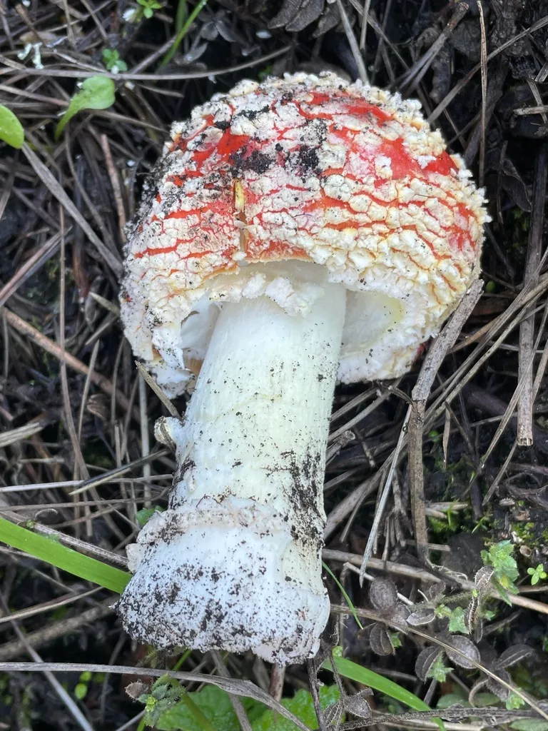
Volva at the base — a cup or sack of tissue from which the mushroom emerged. Feel the base of the stem in the soil. If there's a bulbous, sack-like structure, this is a structural feature unique to Amanita and a few relatives. Amanita phalloides (Death Cap) always has a volva, but it's often buried — you have to dig.
Universal veil remnants — warts or patches on the cap are torn remnants of the universal veil that enclosed the young mushroom. They feel powdery or cottony and can be removed. Note: rain washes them off, so a smooth Amanita cap is still Amanita.
Partial veil / ring (annulus) — a membranous skirt on the upper stem. Texture ranges from fragile and membranous (A. phalloides) to thick and grooved (A. caesarea). Its presence, position, and texture help key species.
Odor in Amanita:
Here's the honest limitation — the most dangerous Amanita species (phalloides, ocreata, bisporigera) smell relatively unremarkable when fresh. A. phalloides has been described as having a faint honey-like or rose-like smell that is mild and not alarming. Older specimens can smell unpleasant, but that's degradation, not a species-specific warning.
This is a critical point: you cannot smell your way out of an Amanita poisoning. The genus that most needs multi-feature identification is also the genus where odor is least helpful as a primary diagnostic. The structural texture features — volva, ring, cap warts — are what matter in Amanita.
What odor does do: some edible Amanita species (A. caesarea, A. rubescens) have recognizable scent profiles to experienced handlers. But beginners should not use odor as a confidence-builder with this genus. Use texture, structural anatomy, spore print (white), habitat, and ideally expert verification.
7. Chemical Spot Tests as an Extension of Sensory ID
Sensory testing — smell, feel, taste — is field chemistry without equipment. Chemical spot tests extend that same principle using reagents that trigger visible color reactions based on specific compounds present in the tissue. They bridge field work and laboratory mycology.
7.1 KOH Reaction and What It Reveals
Potassium hydroxide (KOH) solution — typically 10% concentration — is the most widely used chemical reagent in field and herbarium mycology. A drop applied to cap surface, flesh, or spores triggers color changes based on the alkaline reaction with specific phenolic and pigment compounds in the tissue.
How to apply: Drop a small amount of KOH solution onto the fresh-cut flesh or cap surface. Observe for 30–60 seconds. Note the immediate reaction and any color development.
What reactions mean:
| Reaction | What It Indicates | Example |
|---|---|---|
| Yellow to orange | Presence of specific pigments, Russula group reactions | Russula species subgrouping |
| Red to purplish-red | Phenolic compounds, some Agaricus | Helps confirm A. xanthodermus |
| No reaction | Absence of reactive compounds | Also diagnostically useful |
| Dark brown to black | Specific tannin-like compounds | Some Cortinarius |
For the Agaricus group, KOH on the cap surface of A. xanthodermus produces a bright yellow reaction — the same yellowing you see from mechanical bruising, but faster and more controlled. Applied to A. campestris or A. bisporus relatives, the reaction is absent or very faint. This gives you a third independent test alongside smell and bruising reaction.
In Russula, KOH reactions help divide large species complexes. The reaction on gill tissue can distinguish between acrid and mild species groupings, and on the cap cuticle it differentiates pigment types that aren't visible to the naked eye.
KOH is also used on dried herbarium specimens — the smell and texture of a dried mushroom are lost, but the KOH reaction is preserved and reproducible. This is why chemical features are included in formal taxonomic descriptions.
Practical field use: Small KOH vials are cheap and stable. Serious foragers carry them alongside hand lenses. A single drop is all you need, and the reaction is immediate.
7.2 Melzer's Reagent — Spore Surface Texture Check
Melzer's reagent (iodine solution with chloral hydrate and potassium iodide) is used primarily to examine spore reaction and to reveal microscopic texture features that aren't visible otherwise. It's less common as a field tool and more standard in microscopy work, but it extends the texture concept down to the cellular level.
Amyloid vs. Inamyloid reaction:
- Amyloid: spores or tissue turn blue-black with Melzer's — indicates the presence of specific polysaccharide compounds (amylose-like). Example: Russula and Lactarius spores are amyloid.
- Dextrinoid: tissue turns reddish-brown. Example: some Amanita tissue.
- Inamyloid: no color change. Most spores fall here.
This matters because amyloid/inamyloid status is a hard feature used in formal taxonomy and in keys when other features are ambiguous. Two species that look identical, smell similar, and have similar texture can sometimes be separated definitively by spore reaction to Melzer's.
Cystidia texture under microscopy: Melzer's also helps visualize cystidia — specialized cells on gill edges or surfaces — whose texture and shape are genus-defining. In Inocybe, for instance, the shape of cystidia (thick-walled, thin-walled, crystalline-encrusted) is used to divide species groups that are otherwise visually identical. This is texture analysis at the microscopic scale — the same principle as field texture testing, extended inward.
Spore ornamentation: Under microscopy with appropriate staining, spore surface texture — smooth, warted, ridged, reticulate — is a species-level feature. Russula and Lactarius spores have ornate surface patterns under SEM that correlate with species groupings. The texture of a surface invisible to the naked eye ends up being taxonomically decisive.
The practical takeaway for field foragers: you don't need Melzer's to identify most edible species safely. But understanding that texture exists at multiple scales — cap surface, flesh, gills, cells, spores — explains why professional mycologists can confirm identifications that field workers can only narrow down. Chemical and microscopic tools are extensions of the same sensory logic you apply with your hands and nose, operating at higher resolution.
8. Safety: When Odor and Texture Prevent Poisoning
All of the sensory methodology described in this article ultimately serves one practical purpose: not dying, or at minimum, not spending a night in gastric crisis. This section deals directly with safety — the specific cases where smell and texture are the last line of defense, and the cases where they offer false reassurance.
8.1 Toxic Lookalikes Separated by Smell/Texture
Several of the most dangerous species confusion pairs in foraging are cleanly resolved by sensory testing. These are not edge cases — they represent the most common poisoning scenarios in North America and Europe.
Confusion Pair 1: Giant Puffball (Calvatia gigantea) vs. Egg-stage Amanita
Young Amanita species — including A. phalloides and A. bisporigera — emerge from the soil as white eggs that look externally similar to small puffballs. Foragers have died collecting what they believed were puffballs but were actually Amanita eggs.
The test: cut the specimen in half vertically from top to base.
- True puffball: pure white, homogeneous flesh throughout, no internal structure
- Amanita egg: a cross-section reveals the outline of the future cap, gills, and stem inside the universal veil — a distinct silhouette visible in the white flesh
This is a texture/structure test, not smell-based. But it is absolute. A puffball is solid white throughout with no interior differentiation. If you see any internal structure or outline, it is not a puffball. Never eat an uncut white round object.
Confusion Pair 2: Honey Fungus (Armillaria species) vs. Deadly Galerina marginata
This pair kills people every year. Both grow in clusters on or near wood, have brown caps, ring on stem, and buff-brown gills. Galerina marginata contains the same amatoxins as Amanita phalloides.
Key sensory differences:
- Armillaria: caps typically honey-brown with small dark scales at center (texture). Smell is pleasant, mushroomy, sometimes slightly sweet-honey note. Ring is white to yellowish, membranous.
- Galerina marginata: cap is hygrophanous — pale tan and water-stained when drying. Smooth, no scales. Ring is thin, fragile, often collapses against the stem. Smell is mild, unremarkable, sometimes slightly mealy.
The hygrophanous texture of Galerina — that characteristic two-toned water-stained cap — is the fastest visual-texture cue. Combined with the absence of scales and the fragile ring, it separates the two. The critical rule: never eat a cluster of brown-capped wood-growing mushrooms unless you can confirm every single specimen in the cluster. Galerina and Armillaria frequently fruit in mixed clusters at the same site.
Confusion Pair 3: Penny Bun / Porcini (Boletus edulis) vs. Satan's Bolete (Rubroboletus satanas)
Both are large, fleshy boletes with thick stems. Rubroboletus satanas causes severe GI poisoning.
Sensory differentiation:
- B. edulis: pores white to yellow-green, flesh white and does not change color when cut, mild pleasant odor
- R. satanas: pores red to orange-red (visual, but vivid), flesh turns blue immediately when cut — a dramatic and instantaneous color change caused by an enzymatic oxidation reaction. Unpleasant odor, described as carrion-like when mature.
The bluing reaction is a texture-adjacent test — it's a chemical reaction triggered by cutting, observable in seconds. In boletes, flesh that blues rapidly after cutting is a warning signal. Not all bluing boletes are toxic (Gyroporus cyanescens blues dramatically and is edible), but bluing combined with red pores is a reliable danger signal.
8.2 Amatoxin Species — Why They Smell "Normal"
Amatoxins — specifically alpha-amanitin — are bicyclic octapeptides that inhibit RNA polymerase II in human cells, causing progressive liver and kidney failure with a characteristically delayed onset of 6–24 hours after eating. They are produced by Amanita phalloides, A. ocreata, A. bisporigera, A. virosa, and some Galerina and Lepiota species.
The critical safety fact about amatoxin species: they do not smell dangerous.
A. phalloides in good condition has been described as having a faintly pleasant honey or rose-like smell. It does not smell bitter, chemical, or "off." The flesh has a normal, unremarkable texture. It does not taste bad. These mushrooms have killed experienced foragers in addition to beginners, in part because the organoleptic profile provides no warning.
This is the most important safety message in this entire article: absence of bad smell ≠ safe to eat.
What sensory features can help with amatoxins:
- Structural texture: volva at stem base is always present in Amanita — dig to find it
- Ring texture: membranous, hanging ring on upper stem
- Gill texture: free gills (not attached to stem), white throughout life
- Spore print: white (though this takes hours to develop)
None of these are smell-based. All are texture or structural. The safety protocol for Amanita is anatomy-based, not chemistry-based, because the chemistry provides no signal.
For Galerina, the hygrophanous cap texture is the sensory cue — but it requires knowing what hygrophanous looks like, which requires handling specimens, not reading about them.
The honest limitation of sensory ID in amatoxin species:
Smell and texture are powerful tools for most dangerous lookalike pairs. For amatoxins specifically, they are insufficient without structural anatomy. This is not an argument against sensory ID — it's an argument for understanding which tool applies to which problem.
8.3 Edibility Determination — Smell/Texture as Final Confirmation Step
A practical framework for field edibility determination using sensory ID, applied after initial visual identification:
Step 1 — Visual ID to genus/species candidate Cap color, shape, gill attachment, habitat, spore print (if time allows). This gets you to a short list.
Step 2 — Odor test Smell fresh, immediately after picking. Smell again after cutting the flesh. Smell the stem base separately. Note: pleasant, mealy, chemical, fishy, or fruity. Compare against known profiles for your candidate species.
Step 3 — Cap surface texture Viscid, dry, scaly, hygrophanous, smooth. Rules in or out genera immediately.
Step 4 — Flesh texture Brittle or fibrous? Cut stem lengthwise — hollow, solid, or stuffed? Does flesh change color when cut?
Step 5 — Gill/pore texture True gills or ridges? Waxy, brittle, blade-like? Latex present? What color, what behavior on air?
Step 6 — Structural anatomy (critical for Amanita and Lepiota) Dig the stem base. Volva present? Ring present, where, what texture?
Step 7 — Cross-check against known dangerous lookalikes in your region This is not optional. A correct positive ID is insufficient if you haven't actively ruled out the dangerous lookalike that shares most features.
Step 8 — Eat a small quantity first if new to you Even correctly identified edible species can cause idiosyncratic reactions in some individuals. Armillaria, Morchella, and many others cause reactions in a small percentage of people even when properly cooked.
The role of smell and texture in this framework: they cover steps 2 through 5 — four of eight steps. They do the heavy lifting in the middle of the identification process, where visual features overlap between species and structural anatomy alone isn't enough. Used correctly, they transform an uncertain visual match into a confident multi-feature confirmation.
Mushroom identification is ultimately a discipline of accumulated pattern recognition — you build a library of sensory experiences that your brain matches against new specimens faster and more accurately over time. The chemistry of odor and the biology of texture are not abstract concepts. They are things you smell and feel in the field, and they are the difference between a confident harvest and a dangerous guess.
Sign in to leave a comment and join the discussion.
 Guide
GuideMedicinal mushroom identification means accurately recognizing fungi with proven health-supporting compounds —and distinguishing them from toxic lookalikes....
 Guide
GuideHow Many Mushroom Gummies Should I Eat: A Practical Guide Straight answer: how many mushroom gummies should i eat depends entirely on what's actually in the...
 Guide
GuideI've spent the last six months running functional mushroom gummies through the same scrutiny I bring to wild specimens, checking certificates of analysis,...