Can Two Mushrooms Look Identical but Differ in Toxicity?

Nathan Cole
Mycologist · Author · Fungi Expert
Updated
Apr 30, 2026
Can Two Mushrooms Look Identical But Differ in Toxicity? Absolutely, and the gap between dinner and a liver transplant can come down to a single feature you didn't check. I've sat with families in ICU waiting rooms after a foraged "puffball" turned out to be a button-stage Amanita phalloides. The cap was white. The gills were pale. The forager was certain. The amatoxins didn't care.
In my four decades walking oak stands from the Cascades down through coastal California and across the southern Appalachians, I've watched the same lethal pairs trip up beginners and old hands alike. Galerina marginata on a mossy log next to Armillaria mellea. Chlorophyllum molybdites in a suburban lawn next to a true parasol. Visual identity is not biological identity. Here's the protocol I teach, the pairs that kill, and the line between a good harvest and a 1-800-222-1222 call.
Why Identical-Looking Mushrooms Are a Life-or-Death Question
Two species can share cap color, gill spacing, stipe length, and growth habit and still belong to entirely different toxin classes. Amanita phalloides and a young Agaricus campestris can look like siblings to an untrained eye. One contains α-amanitin. The other contains breakfast.
Amatoxin doesn't taste bitter. It doesn't smell off. It survives boiling, drying, freezing, and sautéing in butter. According to the toxicology committee at the North American Mycological Association, A. phalloides and its close relatives A. bisporigera, A. virosa, and A. ocreata account for the overwhelming majority of fatal mushroom poisonings in North America.
Latency is the cruelest part. Symptoms hit 6 to 24 hours after the meal, by which point the toxin has already started shutting down RNA polymerase II in your hepatocytes. By the time the GI phase passes and you feel "better" on day two, your liver is failing in silence.
If you've eaten a foraged mushroom and feel sick, call Poison Control at 1-800-222-1222 immediately. Don't wait for confirmation, and don't try to identify the species before you call. The dangerous lookalikes here include A. bisporigera in eastern hardwoods and A. ocreata under California oaks, both of which mimic young Agaricus in cap and gill stage.
Who Should Read This Before Their Next Forage
This guide is written for the home forager, the homesteader, the chef sourcing wild ingredients, and the parent whose kid pulled something out of the backyard mulch. It's also for the new mycology student who's read Arora's Mushrooms Demystified twice and is now looking at a real mushroom in real dirt and feeling less sure than the book made it sound.
If you're an ER physician or a poisons specialist, the protocol section assumes you'll cross-reference with the latest Clinical Toxicology literature on silibinin and N-acetylcysteine dosing. This is not a clinical reference. It's a forager's gate-check.
What this guide is not: a substitute for hands-on training with a certified mycologist. The North American Mycological Association maintains a list of regional clubs. Find one. Walk with them. No article, mine included, replaces a person who has held 10,000 mushrooms.
Before any wild mushroom you've identified hits a plate, the rule is simple: get it cleared in person by a certified mycologist, and keep 1-800-222-1222 in your phone.
The Core Protocol: Spore Print, Volva Check, Substrate, KOH
Every confident wild-mushroom ID I've ever signed off on passes through four gates. Skip one, and you've stopped doing mycology and started gambling. Run them in this order, every time, on every specimen, even the ones you "already know."
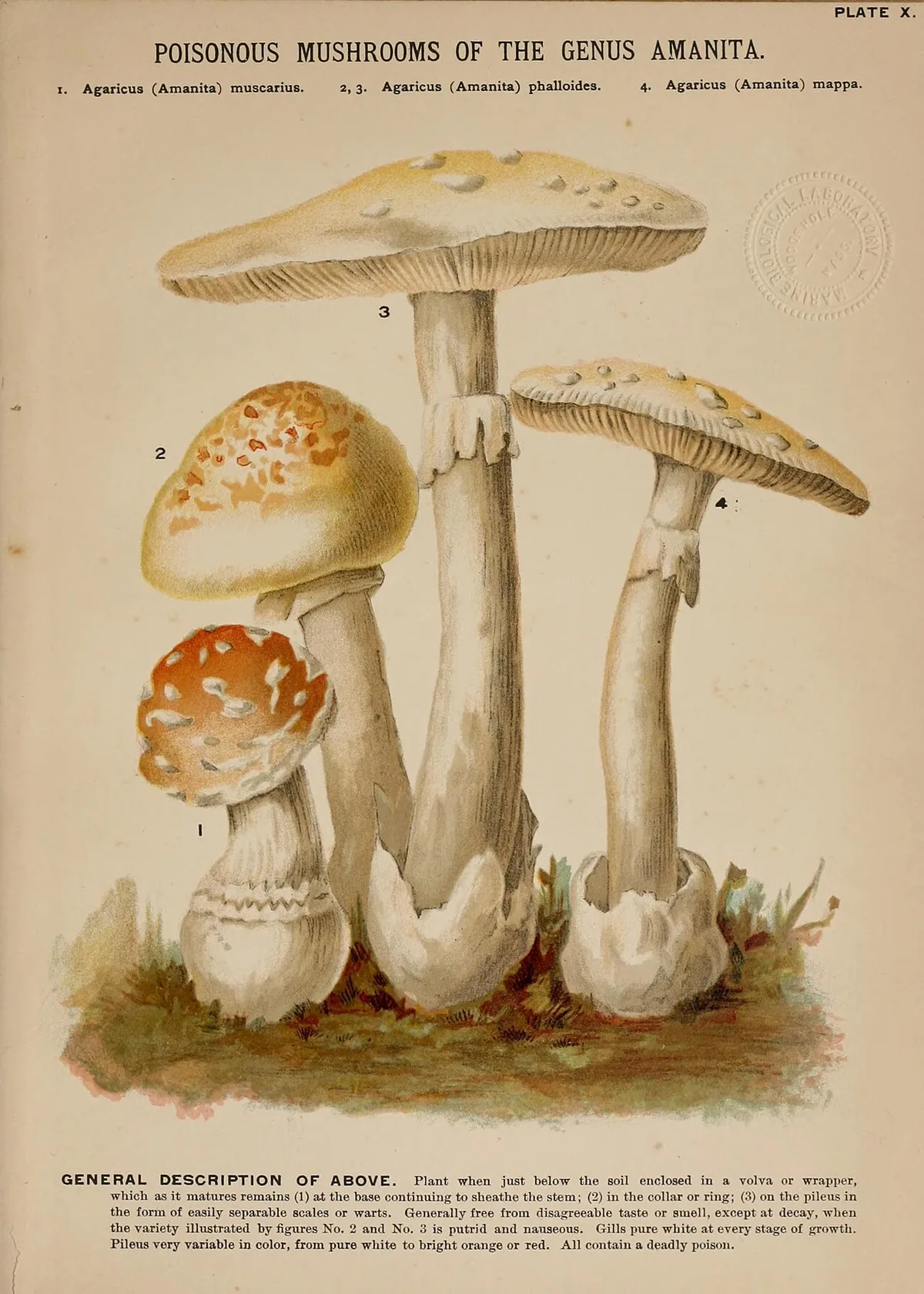
Source Credit: https://commons.wikimedia.org/wiki/File:Mushrooms_of_America_(Plate_X)_(8596720785).jpg
Gate One: Spore Print
Cut the cap. Lay it gills-down on half white paper, half black paper. Cover with a glass. Wait two to twelve hours.
Spore color separates entire genera. White spores point you toward Amanita, Pleurotus, Tricholoma, Armillaria. Pink points to Volvariella and Pluteus. Chocolate-brown points to Agaricus. Rusty-brown points to Galerina and Cortinarius. Bright green points to Chlorophyllum molybdites and nothing edible.
If a foraged "honey mushroom" drops a rusty-brown print instead of white, you don't have Armillaria mellea. You have Galerina marginata, and dinner is cancelled. That single substitution accounts for amatoxin cases I've reviewed from Oregon to Pennsylvania.
Gate Two: Volva Check
Dig the entire base out of the soil. Don't twist, don't snap, don't trim in the field. A sacklike cup at the base, the volva, is the calling card of the Amanita genus. Several of those species (A. phalloides, A. bisporigera, A. virosa, A. ocreata) will kill you.
Half the fatal A. phalloides cases I've reviewed shared one detail: the forager cut the stipe at ground level and never saw the volva.
Gate Three: Substrate
Is it growing from soil in association with a tree? Is it growing from wood? Buried wood? Dung? Substrate narrows the species set fast. Cantharellus formosus and C. californicus are mycorrhizal with conifers and oaks; they emerge from soil as singletons or loose troops. Omphalotus olearius and O. olivascens cluster from buried hardwood roots and stumps. If your "chanterelle" is in a tight cluster on wood, it isn't a chanterelle, and the illudin S in Omphalotus will have you vomiting within two hours.
Gate Four: KOH and Chemistry
A drop of 3 to 10 percent potassium hydroxide on the cap surface of A. phalloides turns yellow. Iron sulfate (FeSO4) helps section Russula, including the rhabdomyolysis-causing R. subnigricans. Melzer's reagent reveals amyloid spore reactions under the scope. The Meixner test (newspaper plus concentrated HCl) gives a presumptive amatoxin reaction, useful as a screen, never as a green light.
If you can't run these gates on a specimen, you don't eat the specimen. Confirmation goes through a certified mycologist, and any suspected ingestion goes through Poison Control at 1-800-222-1222.
The Five Pairs That Kill: A. phalloides/Agaricus, A. bisporigera/Volvariella, Galerina/Armillaria, Gyromitra/Morchella, Cortinarius/Edible Webcaps
These are the pairs I drill into every workshop. Memorize the sharpest separator for each.
Amanita phalloides and Agaricus campestris
The death cap fruits in PNW oak landscaping from late September through January, peaking after the first heavy rains. In California oak woodlands I've seen it from December into March. Agaricus campestris, the meadow mushroom, fruits in pasture and lawn from late summer through fall. Both can present a pale cap and white-ish gills as buttons.
Sharpest separator: free white gills that stay white into maturity, plus a sacklike volva at the base, equals Amanita. Pink-to-chocolate maturing gills with no volva, ever, equals Agaricus. Run a spore print: white kills, brown does not.
Amanita bisporigera and Volvariella volvacea
A. bisporigera, the eastern destroying angel, fruits across Appalachian hardwoods July through October, often under oak and beech. The paddy straw mushroom, Volvariella volvacea, is harvested commercially across Southeast Asia and is sometimes foraged on rice straw and compost in the southern US.
Sharpest separator: spore print. Volvariella drops salmon-pink spores. A. bisporigera drops pure white. Both have a volva. The print is the line.
Galerina marginata and Armillaria mellea
Galerina marginata is a small brown mushroom with rusty-brown spores and α-amanitin in its tissue. It fruits in spring and fall on conifer wood, often on moss-covered logs. Armillaria mellea, the honey mushroom, is a prized edible with white spores and rhizomorph cords running under the bark of host hardwoods. Both can fruit inches apart in October.
Sharpest separator: spore print color. White equals Armillaria. Rusty-brown equals dialysis or worse. If you can't tell them apart, don't eat either, and bring a specimen to a certified mycologist before you ever try.
Gyromitra esculenta and Morchella esculenta
True morels (M. esculenta, M. americana, M. importuna) are hollow from the tip of the cap to the base of the stipe, with a pitted, ridged cap that's fully fused to the stipe. Gyromitra esculenta, the false morel, is brain-like, lobed, with internal chambers stuffed with cottony tissue. Fruiting overlaps in April and May.
The toxin is gyromitrin, which converts to monomethylhydrazine (MMH) in the gut. MMH is hepatotoxic, neurotoxic, and a known carcinogen. Parboiling does not reliably remove it, and the steam itself is dangerous to inhale. Verpa bohemica, the early false morel, complicates things further with a cap attached only at the apex. I do not eat Gyromitra. I do not recommend anyone eat Gyromitra.
Cortinarius rubellus and Edible Webcaps
Cortinarius rubellus and C. orellanus carry orellanine, a renal toxin with a 2 to 14 day latency. The patient often doesn't connect the meal to the kidney failure that follows. There is no edible Cortinarius worth the risk for a non-specialist. Treat the entire genus as off-limits unless you're working under a research mycologist with microscopy. If exposure is suspected, call 1-800-222-1222 immediately, even days after the meal.
Amatoxin Mechanism and the 6–24 Hour Latency Trap
Amatoxins are bicyclic octapeptides. The dominant lethal compound, α-amanitin, binds RNA polymerase II in eukaryotic cells and shuts down protein synthesis. Hepatocytes, with their high metabolic turnover, die first. Renal tubular cells follow.
The lethal dose is roughly 0.1 mg per kg of body weight. A single mature A. phalloides cap can carry more than that. Boiling does not break the toxin down. Drying does not break it down. Cooking with butter, wine, garlic, and forty years of grandmotherly tradition does not break it down.
The clinical course runs in three phases. Phase one is GI distress at 6 to 24 hours: vomiting, watery diarrhea, abdominal cramping. Phase two is the deceptive "honeymoon" at 24 to 72 hours, where the patient feels better and ER teams sometimes discharge them. Phase three is hepatic and renal failure at 3 to 6 days, with mortality north of 10 percent even with aggressive silibinin (Legalon SIL), N-acetylcysteine, IV fluids, and transplant evaluation.
If you suspect amatoxin ingestion, the call goes out before the second wave of vomiting stops. Poison Control: 1-800-222-1222. Bring the remaining mushrooms, the cooking water, and any vomitus to the hospital in sealed paper bags (never plastic, which sweats and degrades the tissue). A working mycologist on the NAMA consult list can confirm A. phalloides or A. bisporigera from one intact specimen faster than any laboratory test.
Contraindications: When You Must Not Trust the Field ID
There are conditions under which my own field call gets overridden by the rules below. If any of these apply, the mushroom doesn't go in the pan. Period.
App-Only Identification (iNaturalist, Picture Mushroom)
Photo-recognition apps are useful for narrowing a genus on a hike. They are not a clearance to eat. iNaturalist's research-grade tag means two humans agreed on a photo, not that the mushroom in your basket is the same species as the one in the photo.
I've seen confident phone screens label Galerina marginata as Armillaria mellea and Chlorophyllum molybdites as Macrolepiota procera. Both errors send people to the ER, and the Galerina miss is potentially fatal. Use apps to start the conversation, not to end it.
Grandmother Always Picked These Folk Knowledge
Folk identification works until the climate, the trees, or the species range shifts. Amanita phalloides didn't exist in North American oak woodlands a century ago. It rode in on imported European nursery stock and has been documented expanding under coast live oak from Vancouver Island to San Diego since the 1970s.
If your grandmother foraged in Krakow, her rules don't transfer to coastal California. Regional variance is not a footnote, it's the whole game. The Mycological Society of San Francisco has documented multiple amatoxin fatalities in immigrant families who ate what looked like the Volvariella volvacea of home and turned out to be A. phalloides.
Cooking, Boiling, and Drying as Detoxification
Heat does not deactivate amatoxin. It does not deactivate orellanine. It does not reliably deactivate gyromitrin, and the volatile MMH that boils off is itself toxic to anyone in the kitchen.
The only toxin classes that respond to heat are certain hemolysins in Amanita rubescens and the heat-labile compounds in Morchella species, and even there, raw or undercooked specimens cause GI distress. If a mushroom needs heat to be edible, that's a feature of the species, not a workaround for a misidentification. Any "I cooked it well, it should be fine" reasoning is the kind of red flag that ends in a call to 1-800-222-1222.
Misapplications I've Seen Send Foragers to the ER
Three pairs account for the bulk of the calls I've taken from emergency departments. Each one has a single feature that would have prevented the ingestion if anyone had checked.
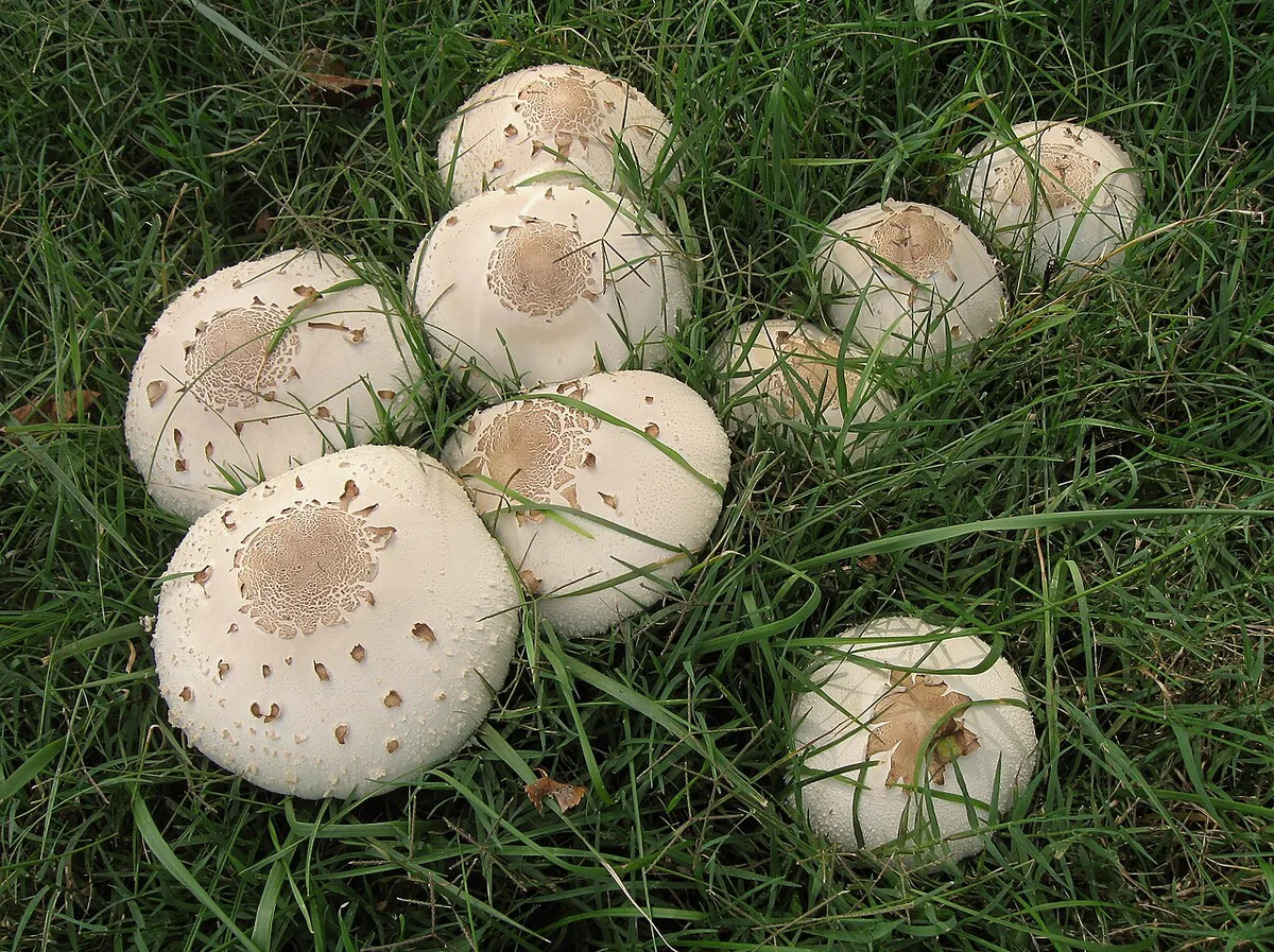
Source Credit: https://commons.wikimedia.org/wiki/File:2011-09-14_Chlorophyllum_molybdites_169974_cropped.jpg
Chlorophyllum molybdites Mistaken for Macrolepiota procera
Chlorophyllum molybdites, the green-spored parasol, is the single most-reported toxic mushroom in North America by call volume to NAMA's poison case registry. It fruits in fairy rings on suburban lawns from late summer through fall, scaly cap, ringed stipe, the whole parasol look.
The true parasol, Macrolepiota procera, is a fine European edible. Same scaly cap, same elegant stipe, same ring. The separator is the spore print: white in Macrolepiota, dull olive-green in Chlorophyllum. Make the print. Every time. The toxin is a GI irritant, rarely fatal in adults, but devastating in children and dogs, and Poison Control should be on the line before the ride to the ER.
Omphalotus olearius Mistaken for Cantharellus cibarius
I've pulled chanterelles from the same Douglas fir slope in the Olympics every September for thirty years. Cantharellus formosus there, C. cibarius in the East, C. californicus under coast live oak. All three have false gills, forked blunt ridges, not knife-edged lamellae.
Omphalotus olearius (East and Mediterranean) and O. olivascens (West Coast) cluster from buried hardwood roots, often around oak and eucalyptus stumps. They have true sharp gills running down a distinct stipe, deeper orange tones throughout, and bioluminesce faintly in the dark when fresh. The toxin (illudin S) causes violent GI illness within two hours. Hygrophoropsis aurantiaca, the false chanterelle, is a separate softer-fleshed lookalike on woody debris, also worth knowing. Run the gill check before the pan ever heats.
Lepiota brunneoincarnata Mistaken for Small Agaricus
Lepiota brunneoincarnata and its close cousins L. josserandii and L. subincarnata carry the same α-amanitin that makes A. phalloides lethal. They're small, brown-scaled, and easy to confuse with a young Agaricus campestris in lawn or park settings.
The separator is the spore print and the gills. Lepiota drops white spores and keeps free white gills into maturity. Agaricus drops chocolate-brown spores and develops pink-to-brown gills. If a small "field mushroom" prints white, stop. The genus Lepiota, with rare exceptions, is a no-eat list for non-specialists. Confirm with a certified mycologist before any consumption decision.
Edge Cases: A. smithiana, Paxillus involutus, Tricholoma equestre, Russula subnigricans
These four species teach a hard lesson. Edibility lists in older field guides are not gospel, and species reclassification plus new toxicology research keep moving the line.
Amanita smithiana fruits in PNW conifer forests under western hemlock and Douglas fir from September through November, often in the same season and habitat as Tricholoma magnivelare, the prized matsutake. The matsutake has an unmistakable cinnamon-spicy odor. A. smithiana doesn't, and it carries an allenic norleucine derivative that triggers acute renal failure 2 to 6 days after ingestion. Smell every matsutake before it goes in the basket.
Paxillus involutus, the brown roll-rim, was eaten across Eastern Europe for generations. We now know it triggers an immune-mediated hemolysis that builds with repeated exposure. People who ate it for thirty years died from a single later meal. Avoid it.
Tricholoma equestre, the man on horseback, was a beloved European edible until a cluster of rhabdomyolysis fatalities documented in Clinical Toxicology in the early 2000s reset the consensus. Russula subnigricans in East Asia and parts of the southeastern US causes the same muscle-tissue breakdown, with peer-reviewed case reports in Toxicon.
If a "classic edible" sits on the borderline of a recent journal case report, it's not on my plate. Foraging is not a tradition I'm willing to defend with my kidneys. When in doubt, certified mycologist first, then 1-800-222-1222 if anything goes sideways.
When to Escalate: Symptom Timeline and the Call to 1-800-222-1222
The single most important number in this guide is 1-800-222-1222. That's US Poison Control, 24 hours a day, free, no insurance question. Call before you Google. Call before you wait it out. Call before the second symptom shows up.
Use this timeline as your trigger map. If a foraged-mushroom meal precedes any of these, the call goes out.
| Time after meal | Symptom pattern | Likely toxin class | Action |
|---|---|---|---|
| 30 min to 2 hr | GI distress, sweating, salivation | Muscarine (Inocybe, Clitocybe), illudin (Omphalotus), GI irritants (Chlorophyllum) | Call 1-800-222-1222, bring specimens |
| 2 to 6 hr | Vomiting, cramping, no fever | GI irritants (Chlorophyllum molybdites, Agaricus xanthodermus) | Hydrate, call Poison Control |
| 6 to 24 hr | Severe vomiting, watery diarrhea | Amatoxin (A. phalloides, A. bisporigera, Galerina, Lepiota brunneoincarnata), gyromitrin (Gyromitra) | ER NOW, bring all remaining mushrooms |
| 24 to 72 hr | Symptoms fade, then jaundice | Amatoxin phase 2 (deceptive honeymoon) | ER admission, hepatology consult |
| 2 to 14 days | Reduced urine, flank pain | Orellanine (Cortinarius rubellus, C. orellanus), allenic norleucine (A. smithiana) | ER, nephrology, dialysis prep |
Bring the mushroom. Bring the cooking pot. Bring the trash bag with the trimmings. A working mycologist on the hospital consult list can confirm the species from one intact specimen, and that confirmation drives the treatment protocol. Silibinin (Legalon SIL) and N-acetylcysteine work better the earlier they start.
If you're in the field and someone in your group eats an unknown mushroom, don't wait for symptoms. Call Poison Control immediately. Save a whole specimen, base and all, in a paper bag (never plastic, which sweats and degrades the tissue).
References: NAMA, Poison Control, Field Guides, and Peer-Reviewed Toxicology
The sources below are the ones I keep on the shelf and consult before I sign off on any identification or write any guide like this one.
Poison Control: 1-800-222-1222, available 24/7 across the United States. The single first call for any suspected mushroom poisoning.
The North American Mycological Association maintains the toxicology committee that tracks annual case reports and the volunteer identifier list that hospital ERs call when a specimen comes in. Find your regional society through them: Puget Sound Mycological Society, Mycological Society of San Francisco, Cumberland Mycological Society, New York Mycological Society, and dozens more.
Field guides I trust on my shelf: David Arora's Mushrooms Demystified and All That the Rain Promises and More. Kent McKnight's Peterson Field Guide to Mushrooms. Gary Lincoff's Audubon Field Guide. Bessette and colleagues on North American Boletes and Tricholomas of North America. Trudell and Ammirati's Mushrooms of the Pacific Northwest. Michael Kuo's Morels.
For peer-reviewed toxicology and taxonomy, I read Mycologia, Fungal Diversity, Mycoscience, Persoonia, Toxicon, and Clinical Toxicology. The CDC's Morbidity and Mortality Weekly Report has published amatoxin case series that every clinician handling foraged-mushroom cases should know.
There are old mushroom hunters. There are bold mushroom hunters. There are no old, bold mushroom hunters. Make the spore print. Dig the volva. Walk with a club. Call Poison Control at 1-800-222-1222. And before any wild mushroom hits your plate, get it cleared by a certified mycologist in person, not a phone screen.
Sign in to leave a comment and join the discussion.
 Safety
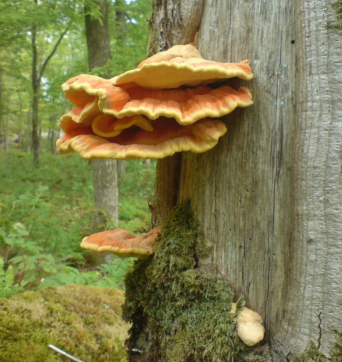
Last September I stood at the base of a fire-scarred white oak in the Blue Ridge foothills and pulled twelve pounds of Laetiporus sulphureus from a single...
 Safety
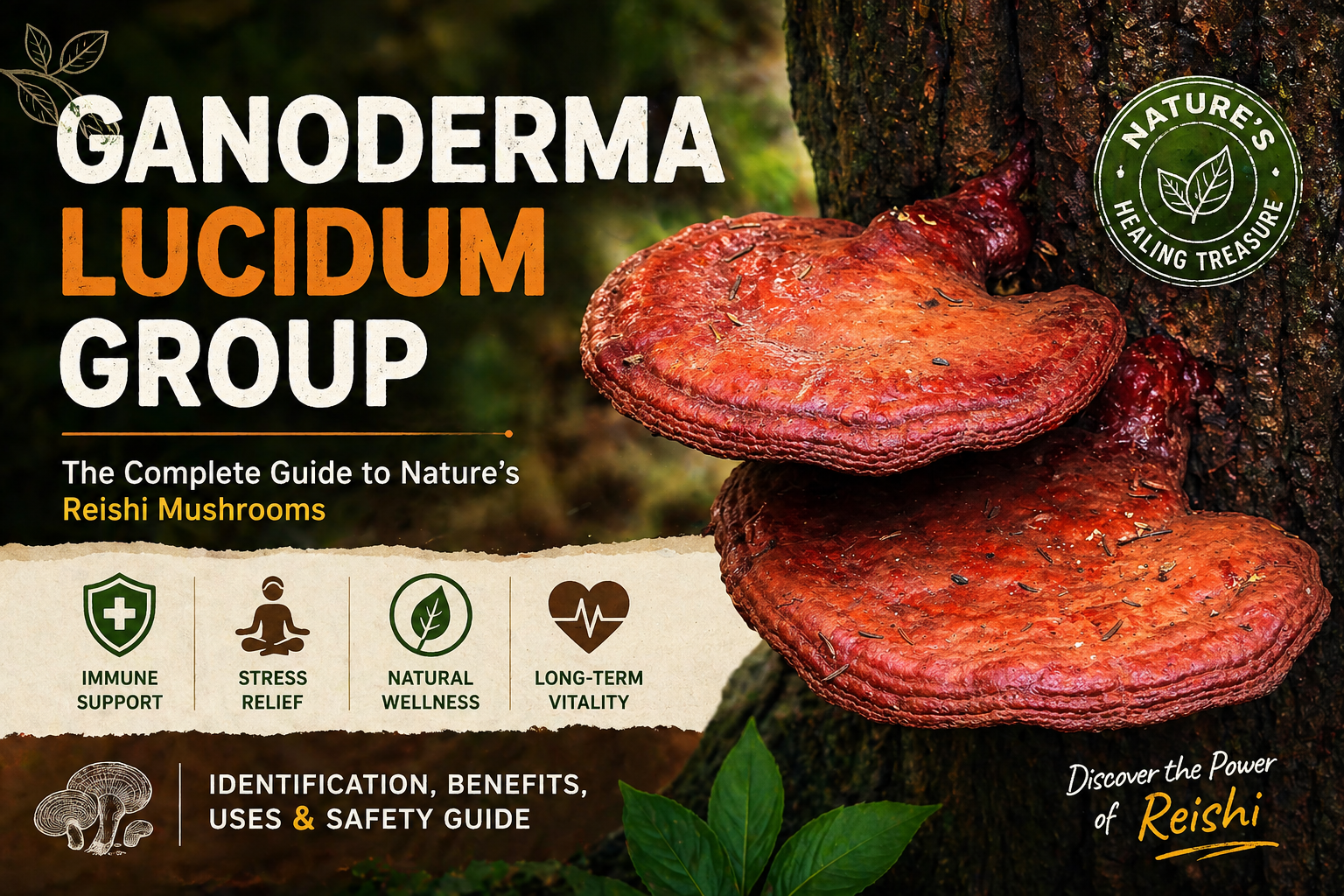
The Ganoderma lucidum group is not a single species. It's a complex of at least a dozen related polypore fungi, including Ganoderma tsugae , Ganoderma...