Can AI Identify Poisonous Mushrooms Safely? Hands-On Review

Nathan Cole
Mycologist · Author · Fungi Expert
Updated
Apr 28, 2026
Can AI Identify Poisonous Mushrooms Safely?: Hands-On Review
After forty years identifying fungi across the Pacific Northwest, Appalachia, and Europe, I'm watching a dangerous pattern take hold: foragers trusting app confirmations of Amanita phalloides (death cap). Can AI identify poisonous mushrooms safely? No, not reliably, and the failure modes are tied to the exact species most likely to kill you. The technology's limits begin at the species level.
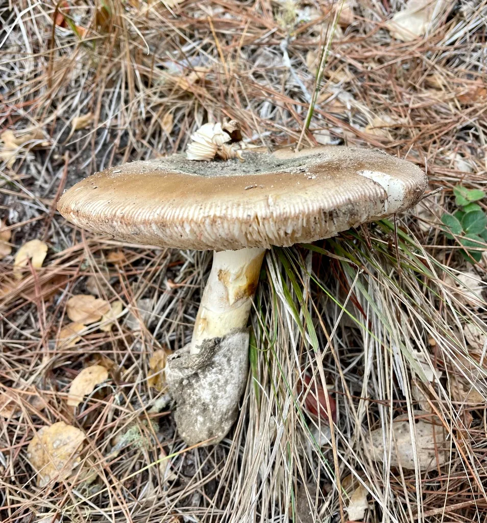
Source Credit: https://www.inaturalist.org/observations/141036614
What "AI Mushroom Identification" Actually Tests and the Identification Gap It Cannot Close
When you photograph a mushroom and submit it to an identification app, the software runs the image through a neural network trained on labeled photographs and returns a probability match. That's the entirety of the operation: pattern-matching pixels against a training dataset.
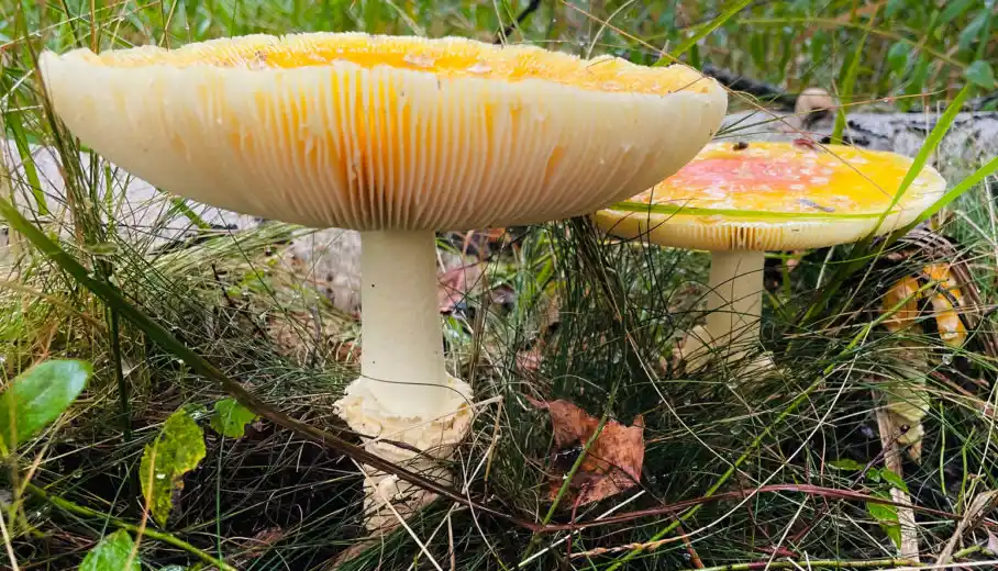
What the software can assess from a photograph: cap color, gross shape, surface texture, approximate gill color when visible, and growth habit. What it cannot assess covers the actual substance of mycological identification. Spore print color. Gill attachment type. The presence or absence of a volva buried at the stipe base. Flesh bruising reactions. Chemical spot tests. Odor. Any microscopic feature. These are precisely the criteria that separate Amanita phalloides from its edible lookalikes, and every single one requires physical interaction with the specimen.
The California Academy of Sciences, which operates the iNaturalist and Seek platforms, states explicitly in its own documentation that these tools are not designed for foraging or consumption decisions. That's the institution behind the best-resourced visual identification platform in existence, saying directly that its own tool isn't safe for this purpose.
A 94% confidence match on a deadly species is not reassuring. It describes the model's uncertainty, not the specimen's edibility. At alpha-amanitin doses of approximately 0.1 mg per kilogram of body weight, a 6% error margin is a mortality margin.
The Three Scenarios Where Foragers Reach for an App Instead of an Expert
The dangerous use cases fall into three patterns, each with a distinct failure mode.
The solo field forager. Someone hiking the Cascades in October finds what looks like Cantharellus cibarius (golden chanterelle) along a Douglas-fir trail. They photograph it, get an 89% chanterelle match, and fill their basket. What the app couldn't assess: whether those orange fruiting bodies have the blunt, forking, decurrent ridges of a true chanterelle or the knife-edge true gills of Omphalotus olivascens (western jack-o'-lantern). Examining gill structure requires a lateral cross-section. No overhead photograph captures it.
The backyard parent. A child brings a white button mushroom in from the lawn. The parent photographs it, gets a positive Agaricus campestris (field mushroom) result, and considers it resolved. The app cannot check whether those gills are pink (young A. campestris, edible) or white (potential Amanita species, potentially lethal). Nor whether a volva is buried at the stipe base. One outcome is a good meal. The other is amatoxin poisoning.
The culturally familiar species. Documented in case series published in Clinical Toxicology, this is the most heartbreaking failure mode. Southeast Asian immigrants in California forage for Volvariella volvacea (paddy straw mushroom), which they know from home. Amanita phalloides growing near ornamental oaks in the Bay Area shares enough visual features that misidentifications have caused multiple fatalities. An AI tool trained primarily on North American and European images compounds rather than resolves this confusion.
The Physical Identification Protocol No App Can Complete
Safe identification is a multi-step physical process. Every step requires handling the specimen. None of it is optional for any species with a potentially lethal lookalike.
Spore Print: The Overnight Test That Separates Galerina marginata from Every Edible Wood-Rotter
Remove the cap. Place it gill-down on a sheet that is half white paper, half black paper. Cover it to prevent air currents. Leave it four to twelve hours, then lift the cap.
A rusty-brown deposit from a honey-colored, wood-growing cluster: that's Galerina marginata, which contains the same alpha-amanitin as A. phalloides. A white deposit from the same-looking cluster: potentially Flammulina velutipes (velvet shank, enokitake), a genuinely good edible. Same habitat. Same season. Same color profile. One destroys the liver. The other is sold in every Asian grocery store. The spore print separates them in one night, and no AI app performs this test.
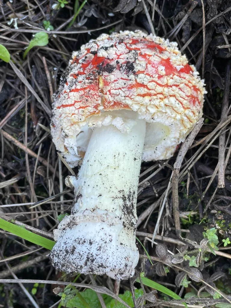
Volva Excavation: The Buried Feature That Distinguishes Amanita phalloides from Every Safe Lookalike
The volva is a cup-like universal veil remnant at the base of the stipe in A. phalloides, A. bisporigera, and A. ocreata, partially or fully buried in the substrate. It is the most important diagnostic feature in the death cap complex, and it is invisible in the vast majority of field photographs.
When I encounter any white-gilled, ring-bearing mushroom in mixed hardwood forest, I dig. I use a knife to expose the full stipe base before anything else. A sack-like volva present: the specimen is an Amanita, and without species-level certainty from expert review, it stays in the ground. An AI app receives whatever angle the photographer chose. Almost always a top-down view of the cap. The volva doesn't appear in that frame. This is not a fixable software problem. It is a physical constraint.
Gill Structure, Bruising Reaction, Odor, and Chemical Spot Tests
Gill attachment requires a lateral cross-section with good lighting: free gills characterize Amanita; blunt, forking, decurrent ridges characterize Cantharellus; knife-edge true gills running down the stipe characterize Omphalotus. These distinctions are not visible in overhead photographs.
Flesh reaction: slice any Agaricus stipe base and watch for ten seconds. Chrome-yellow staining indicates Agaricus xanthodermus, which causes severe gastrointestinal illness. No staining, a pink-to-brown gill progression, and a clean mushroom smell: A. campestris (field mushroom) is the candidate. A. xanthodermus smells of phenol or ink. A. campestris smells like fresh grocery-store button mushrooms. These are physical assessments. Image analysis cannot reach them.
Amanita phalloides, A. bisporigera, and A. ocreata: Where App Confidence Becomes Amatoxin Certainty
These three species, all within Amanita section Phalloideae, account for the overwhelming majority of fatal mushroom poisonings worldwide. The primary toxins are bicyclic octapeptides, chiefly alpha-amanitin, which inhibits RNA polymerase II and halts protein synthesis in hepatic cells. A single cap of A. phalloides can contain 10 to 15 milligrams of alpha-amanitin. The documented lethal dose: approximately 0.1 mg per kilogram of body weight, as established in the toxicological literature compiled in Ammirati, Traquair, and Horgen's Poisonous Mushrooms of the Northern United States and Canada (University of Minnesota Press, 1985). Less than half a cap for a child. Amatoxins are thermostable and water-soluble: cooking, parboiling, drying, and freezing do not neutralize them.
The onset timeline is what makes this poisoning so treacherous. Phase one: a latent period of six to twenty-four hours post-ingestion with no symptoms whatsoever. The patient feels entirely well. Phase two: violent cholera-like gastrointestinal illness begins. Phase three: apparent recovery, a false remission lasting up to seventy-two hours, which leads patients and families to believe the crisis has passed. Phase four: fulminant hepatic failure.
The false remission is the killer. Do not wait for symptoms to return.
If you or anyone near you has eaten a wild mushroom and identification is uncertain, call Poison Control immediately at 1-800-222-1222. That line operates twenty-four hours a day. Provide the full description, the quantity consumed, and the time of ingestion.
A. phalloides was introduced to North America with European oak plantings and is now established across coastal California, fruiting from September through January. A. bisporigera dominates eastern deciduous forests from the Great Lakes to the Gulf Coast, peaking August through October. A. ocreata fruits in winter and early spring along the Pacific Coast under live oak and tan oak.
The Egg-Stage Problem: When a Death Cap Photographs as a Puffball
Before the universal veil ruptures, all three species emerge as smooth, white, egg-shaped structures. From above, in a photograph, they are nearly indistinguishable from edible puffballs (Calvatia and Lycoperdon species). AI has no reliable mechanism to separate them at this developmental stage: both are white, smooth, ovoid, and soil-emerging.
The field test takes thirty seconds. Cut the specimen vertically with a clean knife. A true puffball is uniformly solid white inside. An Amanita egg reveals the silhouette of cap, gills, and stipe already formed within the veil. This requires a knife. No photograph substitutes for it.
The Volvariella volvacea Confusion and the Communities It Has Killed
Volvariella volvacea carries a pink spore print and has no annulus on the stipe. Amanita phalloides has white gills throughout, a distinct membranous ring, and a buried volva. These distinctions are visible to a trained eye in adequate lighting, and completely unreliable in the kinds of photographs AI apps process. Case series in Clinical Toxicology confirm repeated fatalities in California's Southeast Asian immigrant communities from exactly this confusion. NAMA maintains a network of volunteer toxicology consultants, accessible through namyco.org, who can review physical specimens before any foraging decision is finalized.
Galerina marginata: The Log-Growing Amatoxin Source That Defeats Pattern Matching
Galerina marginata is the species that kills experienced foragers who believe they know what they're looking at. Small, honey-colored to tan, hygrophanous (fading noticeably as it dries), bearing a fragile ring that weathers away on older specimens, growing in dense clusters on decaying conifer and hardwood logs from September through December. It contains alpha-amanitin at concentrations comparable to A. phalloides.
So does Flammulina velutipes grow on decaying wood in autumn. So does young Armillaria mellea (honey mushroom). The visual overlap in field conditions, photographed from above in imperfect lighting, is substantial enough that image-based classification cannot reliably separate them. Multiple fatal poisonings in North America have resulted from this exact confusion, documented in Clinical Toxicology case reports.
The separating test is the spore print: G. marginata drops rusty-brown; F. velutipes drops white. That test requires one night and a sheet of paper. No app performs it. Treat every brown, ring-bearing cluster on decaying wood as suspect until a spore print has been completed and, where any uncertainty remains, a certified mycologist has reviewed the physical specimen. Consult a certified mycologist before consuming any wood-rotting species you cannot fully key out through a current field guide combined with spore print confirmation.
Gyromitra esculenta vs. Morchella esculenta: The Spring Misidentification That Gyromitrin Makes Lethal
This is the confusion that kills morel hunters, and it happens every single spring across the Upper Midwest and Pacific Northwest. Both species emerge from sandy soils near conifers when snowbanks are still receding, sometimes from the same hillside in the same week, from March through May.
Morchella esculenta (common true morel) has a cap covered in regular, honeycomb-like pits separated by vertical ridges. The cap is fully fused to the stipe at its base. Cut it vertically: the interior is completely hollow, the cap and stipe forming a single continuous chamber.
Gyromitra esculenta (false morel) has a cap that is irregularly lobed, brain-like, with wrinkled folds that overhang the stipe rather than fusing with it. Cut it vertically: the interior is chambered and irregular, not a clean hollow. From above in a photograph, both species present as brown, crinkled structures emerging from damp conifer soil. That is where image classification fails, and it fails completely.
The toxin is gyromitrin, which hydrolyzes in the body to monomethylhydrazine (MMH), a compound structurally related to rocket propellant hydrazine. MMH disrupts pyridoxal-5-phosphate metabolism, inhibits GABA synthesis, and in severe cases causes methemoglobin formation and hemolytic anemia. Onset runs six to twelve hours.
The widely repeated belief that parboiling and discarding the cooking water makes G. esculenta safe is not reliable. Gyromitrin is partially volatile, so prolonged open-air boiling reduces toxin load, but the reduction is not predictable or consistent, and the vapor produced during boiling is itself toxic to the cook. I do not eat this species under any preparation method, and I advise the same without exception.
If you receive a morel match from an app on a spring-emerging brown cap, that result tells you nothing about whether the cap attachment is correct. Cut the specimen. Check the base. Bring it to your regional mycological society before anything goes in the pan.
Cantharellus cibarius vs. Omphalotus illudens: Why True Gills and False Gills Are Invisible in a Photograph
I mentioned the chanterelle-jack-o'-lantern confusion earlier in the context of the solo field forager, but the structural anatomy here deserves full treatment, because this is where confident, experienced-looking foragers get hurt most often.
Cantharellus cibarius (golden chanterelle) does not have true gills. Beneath the cap are forking, blunt-edged ridges that run down onto the stipe (decurrent) and anastomose, meaning they fork and rejoin in irregular patterns. These ridges are shallow, rounded at their edges, and resist being separated from the cap flesh cleanly.
Omphalotus illudens (jack-o'-lantern mushroom, eastern North America) and its western counterpart O. olivascens have true gills: thin, crowded, knife-edge blades arranged radially beneath the cap. They detach cleanly. The flesh, when you cut through it, is orange throughout. A chanterelle's flesh is white.
In fresh, living Omphalotus specimens, the gills are bioluminescent, producing a faint green glow visible only in complete darkness with dark-adapted eyes. It's a striking feature, but it's not a reliable field test: the luminescence requires fresh tissue, and by the time most foragers examine a collection at home, it has often faded.
The safe protocol has nothing to do with photography. Examine the underside with a hand lens, from the side and below. Blunt, forking, shallow ridges that run onto the stipe: chanterelle. Blade-like true gills, cleanly organized, fully formed, easily separable: put it down. A photograph taken from above, angled at the cap surface, gives you the color profile and nothing of the diagnostic anatomy.
Cortinarius rubellus and the Orellanine Latency Trap: Kidney Failure Three Weeks After the Meal
Of all the failure modes I've described here, the orellanine latency is the one that most completely defeats any identification tool. The toxin in Cortinarius rubellus (deadly webcap) and C. orellanus (fool's webcap) doesn't announce itself for two to three weeks after ingestion. By then, the meal is forgotten, the mushrooms are gone, and the patient presenting with oliguria, flank pain, and rising creatinine is being treated for what appears to be an idiopathic renal event.
I've spoken with clinicians who have seen these cases. The standard intake question, "Did you eat anything unusual recently?", applied on the day of presentation, doesn't reach back three weeks. The diagnosis requires a mycologist in the conversation or a physician who knows to ask specifically about mushroom foraging over the past month.
The mechanism is tubulo-interstitial nephritis. Orellanine and its metabolite orelline inhibit alkaline phosphatase and disrupt cellular protein kinase activity, producing progressive destruction of renal tubular epithelium. There is no antidote. Outcomes range from partial recovery to permanent renal insufficiency requiring dialysis or transplant.
C. rubellus is a rusty orange-brown species fruiting under spruce and pine in northern conifer forests, found in the Cascades, Northern Rockies, and across Scandinavia and the British Isles. The cobweb-like cortina (partial veil remnant) that helps identify the genus disappears with rain and age, leaving older specimens with no veil feature visible at all. An AI app then has nothing to flag.
The practical guidance for the genus is simple: with approximately 2,000 Cortinarius species in North America and Europe, dozens confirmed as orellanine-bearing, and no reliable macroscopic feature separating the lethal from the innocuous at species level, I do not eat any Cortinarius species. If you have any reason to suspect ingestion of a Cortinarius collection from within the past month, call Poison Control at 1-800-222-1222 and document the date and location of the meal.
If You've Already Eaten It: Amatoxin Symptom Timeline, the False Remission Window, and What to Tell Poison Control
As described in the Amanita section above, amatoxin poisoning progresses through four phases over approximately seven days, with the false remission in phase three being the window when patients most commonly decide the crisis has resolved. It hasn't. That apparent recovery is the most dangerous moment in the entire poisoning timeline, because it is when people stop seeking treatment.
The action protocol, regardless of which toxic species is suspected, begins with one call:
Call Poison Control at 1-800-222-1222 immediately. Do not wait for symptoms to appear or for existing symptoms to worsen. When you call, have ready: the time of ingestion, an estimate of how much was eaten, a physical description of the mushroom (cap color and size, gill color, presence of a ring or volva, growth habitat, substrate), and the precise collection location. If any part of the specimen is still available, place it in a paper bag (not plastic, which accelerates decomposition) and refrigerate it. Preserve it for the toxicologist.
Do not induce vomiting and do not administer activated charcoal on your own initiative. Both decisions belong to the Poison Control toxicologist on that call.
At the emergency room, state specifically that amatoxin poisoning is a possibility. Treatment options include N-acetylcysteine; silibinin (the pharmaceutical compound derived from milk thistle, available as Legalon SIL in Europe) has shown benefit in early clinical intervention and may be accessible through a toxicology consult. Liver function panels, coagulation studies, and renal function markers should be monitored through the full first week, including during any period when the patient subjectively feels well.
For suspected Cortinarius ingestion, the same number applies and the same documentation principle holds: the date of the meal may be the only information a treating nephrologist receives.
Consult a certified mycologist before any foraged specimen is consumed. After a suspected ingestion, consult Poison Control and a toxicologist. These are not redundant steps. They are opposite ends of the only protocol that reliably prevents fatalities.
Authoritative Sources, Regional Mycological Societies, and the Only Safe Identification Standard
There is no substitute for expert in-person identification of a physical specimen. That is the standard. Everything below it, including AI apps, photograph-based forums, and beginner field-guide matching, carries risk proportional to its distance from that standard.
Poison Control (United States): 1-800-222-1222. Available twenty-four hours a day, 365 days a year. Bilingual. Free.
North American Mycological Association (NAMA): namyco.org. Maintains a Toxicology Committee, a national network of trained volunteer identifiers, and a club locator connecting foragers to regional societies across North America. NAMA-affiliated clubs hold regular forays and identification sessions where physical specimens are reviewed by experienced mycologists working the same regional flora.
Published field guides:
Arora, David. Mushrooms Demystified, 2nd ed. Ten Speed Press, 1986.
McKnight, Kent H. and Vera B. McKnight. A Field Guide to Mushrooms: North America. Peterson Field Guides, Houghton Mifflin, 1987.
Ammirati, J.F., Traquair, J.A., and Horgen, P.A. Poisonous Mushrooms of the Northern United States and Canada. University of Minnesota Press, 1985.
Benjamin, Denis R. Mushrooms: Poisons and Panaceas. W.H. Freeman, 1995.
Peer-reviewed literature: Mycologia (Mycological Society of America), Clinical Toxicology (Taylor and Francis), Fungal Diversity (Springer), and Mycoscience (Mycological Society of Japan) are the primary sources for taxonomy, toxicology, and clinical case data throughout this article.
Your nearest regional mycological society, found through NAMA's website, offers the most practical resource available to a working forager: someone who has spent years in the same forest types you're walking, who can hold your specimen, run a spore print, cut the base, smell the flesh, and check every feature that a camera cannot capture.
The old saying holds as firmly today as it did before smartphones existed: there are old mushroom hunters, and there are bold mushroom hunters, but there are no old, bold mushroom hunters. AI doesn't change that. It only adds a new way to be bold.
Sign in to leave a comment and join the discussion.
 Guide
GuideMedicinal mushroom identification means accurately recognizing fungi with proven health-supporting compounds —and distinguishing them from toxic lookalikes....
 Guide
GuideHow Many Mushroom Gummies Should I Eat: A Practical Guide Straight answer: how many mushroom gummies should i eat depends entirely on what's actually in the...
 Guide
GuideWhy Odor and Texture Matter in Mushroom ID? Odor and texture are two of the most reliable tools in mushroom identification — and experienced foragers treat...