White Poisonous Mushrooms Identification: A Complete Field Guide to the Species That Kill

Nathan Cole
Mycologist · Author · Fungi Expert
Updated
Apr 20, 2026
White Poisonous Mushrooms: A Complete Field Guide to the Species That Kill
The most dangerous mushrooms in North America are white. Amanita bisporigera — the Destroying Angel — along with Amanita ocreata, Amanita verna, and Amanita virosa account for the majority of fatal mushroom poisonings worldwide. They contain amatoxins — specifically alpha-amanitin — a compound that irreversibly shuts down RNA polymerase II and destroys liver and kidney tissue over days.
There is no universally available antidote. If you've eaten an unidentified white mushroom, stop reading and call Poison Control: 1-800-222-1222 right now.
I've spent over four decades walking forests from the Olympic Peninsula to the Appalachian ridgelines, pulling back leaf litter to find these beautiful, lethal fruitbodies. I've also sat with emergency physicians who've watched patients die from them.
This guide covers every white poisonous mushroom species you're likely to encounter, the toxins inside them, how to identify them by pileus, stipe, volva, annulus, and spore print — and crucially, what they look like next to edible lookalikes. Use it alongside a certified mycologist and David Arora's Mushrooms Demystified. Never use it as your sole identification source.
What Makes a White Mushroom Deadly?
Not all white mushrooms are toxic. But the ones that are — particularly the Amanita Destroying Angels and the Lepiota dapperlings — carry toxins so potent that half a pileus of Amanita bisporigera contains a lethal dose for an adult human. Understanding what those toxins are, how they work, and why the symptoms take so long to appear is the foundation of everything else in this guide.
Amatoxins: Alpha, Beta, Gamma-Amanitin
Amatoxins are bicyclic octapeptides. That sounds abstract until you understand what they actually do inside your body.
Alpha-amanitin is the primary killer. It binds with extraordinary affinity to RNA polymerase II — the enzyme every nucleated cell in your body uses to transcribe DNA into messenger RNA. When alpha-amanitin locks onto it, that process stops. Cells that replicate rapidly die first: intestinal epithelium, hepatocytes in the liver, and renal tubular cells. The liver fails. The kidneys follow. In severe cases with no intervention, the patient dies between day four and day eight.
Beta-amanitin and gamma-amanitin work through the same mechanism, though with slightly lower potency. The three together form the lethal payload carried by Amanita bisporigera, A. ocreata, A. verna, A. virosa, Lepiota subincarnata, Lepiota josserandii, and Conocybe filaris.
What makes amatoxin poisoning particularly cruel is its heat stability. Cooking doesn't destroy these compounds. Neither does drying, freezing, or extended storage. The mushroom can be thoroughly cooked to a pleasing golden brown and still kill you.
| Amatoxin | Primary Source Species | Relative Potency |
|---|---|---|
| Alpha-amanitin | A. bisporigera, A. phalloides | Highest |
| Beta-amanitin | A. bisporigera, Lepiota spp. | High |
| Gamma-amanitin | A. verna, A. virosa | Moderate |
Research published in Mycologia and Fungal Diversity has confirmed amatoxin presence across all Destroying Angel species. Data from Mycoscience on Lepiota subincarnata poisonings in Europe has expanded our understanding of how broadly this toxin class is distributed across fungal families.
Phallotoxins and Virotoxins
Phallotoxins — phalloidin and phalloin — are monocyclic heptapeptides found alongside amatoxins in most Amanita species. They're highly toxic in vitro, disrupting actin polymerization in cells. In practice, however, phallotoxins are poorly absorbed through the GI tract in humans, which is why they're now considered secondary actors in clinical poisoning rather than primary causes of death.
Virotoxins are a third class of cyclopeptide found specifically in Amanita virosa — the European Destroying Angel. They work similarly to phallotoxins in binding actin, and their presence gives A. virosa a somewhat distinct toxicological profile from the American Destroying Angels. In transition zones across the upper Midwest and Northeast where both species can occur, that distinction matters for the hepatologist managing a poisoning case.
Muscarine (Clitocybe Group)
Clitocybe dealbata and Clitocybe rivulosa don't contain amatoxins. Their weapon is muscarine — a quaternary ammonium compound that over-stimulates muscarinic acetylcholine receptors in the parasympathetic nervous system. The clinical result is the SLUDGE syndrome: Salivation, Lacrimation, Urination, Defecation, GI distress, Emesis — typically appearing within 30 minutes to 2 hours of ingestion.
Muscarine poisoning is rarely fatal in healthy adults, but it's dangerous in elderly patients, children, and anyone with cardiac conditions. The small, white, crowded-gilled basidiocarps of C. dealbata show up in grassy lawns, parks, and garden edges — exactly where families forage for Marasmius oreades, the Fairy Ring Mushroom. That habitat overlap has caused serious illness more times than the literature fully reflects.
RNA Polymerase II: Why the Mechanism Kills Slowly
This question comes up every time I speak to a general audience: "Why don't people feel sick immediately?"
Your GI epithelium turns over fast — that's the first wave of symptoms, hitting hard 6 to 24 hours after ingestion. But liver hepatocytes are slower to die. As the body's hepatocyte population collapses, liver function markers — ALT, AST, bilirubin — rise sharply, but the patient often feels better for 12 to 24 hours as the GI crisis subsides. This is Phase III, the false remission. Families have gone home from emergency rooms during this window. Patients have told me they thought they were recovering. They weren't.
By the time fulminant hepatic failure announces itself on day four to six, the intervention window has narrowed dramatically. That latency between ingestion and catastrophic organ failure is the biological reason that amatoxin poisoning carries a case fatality rate of 10–30% even in countries with modern hospital care. The RNA polymerase II mechanism doesn't announce itself — it works silently until the damage is irreversible.
The Primary White Killers — Species by Species
There are roughly eight species you need to know cold before you ever pick a white mushroom in the field. Learn their Latin names. Learn their geography. Learn what tree they grow under.
Photo: This image was created by user Ron Pastorino (Ronpast) at Mushroom Observer, a source for mycological images.You can contact this user here. via Wikimedia Commons, licensed CC BY-SA. Source: https://commons.wikimedia.org/wiki/File:Amanita_ocreata_601832.jpg
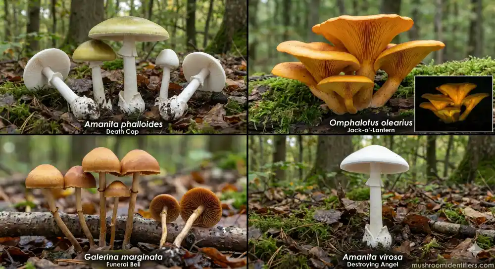
Amanita bisporigera — Destroying Angel (Eastern US)
Amanita bisporigera is the white angel of death east of the Rockies. It's the species most commonly implicated in fatal mushroom poisonings in the eastern United States, and in my estimation, the most dangerous wild fungus on this continent.
Field identification:
- Pileus: Pure white, 5–12 cm, smooth, convex to broadly flat at maturity
- Stipe: White, 8–15 cm, with a distinct pendant annulus hanging from the upper third
- Volva: White, sack-like, partially buried — always dig to find it
- Lamellae: Free, crowded, white — never pinking, never browning
- Spore print: White
- Flesh: White throughout, not staining on cut or with age
The volva is the single most critical feature. Every Destroying Angel emerges from a membranous cup at the base of the stipe. Careless pickers pulling mushrooms without digging miss it entirely — and that omission is often fatal.
Distribution and host trees: Found throughout the eastern US from the Great Lakes to the Gulf Coast, fruiting under mixed hardwoods — Quercus (oak), Betula (birch), and Fagus (beech) — from late summer through fall. I've found this species as far south as Georgia and as far north as Ontario under oak and beech canopy in the Appalachian Mountains.
Toxin load: Among the highest confirmed amatoxin concentrations of any species in the genus. A single cap contains enough alpha-amanitin to kill an adult.
Amanita ocreata — Western Destroying Angel
The West Coast has its own angel. Amanita ocreata is arguably more dangerous to casual foragers because of its early fruiting season and its overlap with a diverse edible mushroom community.
Field identification:
- Pileus: White to ivory, sometimes pale buff at center, 5–12 cm
- Stipe: White, slender, 8–18 cm, with pendant annulus
- Volva: White, membranous, often lobed, partially to mostly buried in duff or soil
- Lamellae: Free, white, crowded
- Spore print: White
- Odor: Faintly unpleasant in age — not a reliable field character
Distribution and host trees: California coastal ranges, Oregon, and Washington — primarily in oak woodland (Quercus agrifolia, Q. garryana) and mixed hardwood-conifer zones. Fruits December through April, overlapping with the spring Cantharellus subalbidus flush in Northern California and Oregon. That timing is a dangerous coincidence, and I'll address it directly in the lookalikes section.
Amanita verna — Fool's Mushroom (Spring Fruiter)
Amanita verna earns its common name honestly. It fruits in spring — April and May across much of North America and Europe — at exactly the moment beginners are excited to get back into the woods after winter.
Field identification:
- Pileus: Pure white, smooth, 4–9 cm, convex to flat
- Stipe: White, 6–12 cm, slender, with a fragile annulus
- Volva: White, well-developed, cupulate
- Lamellae: Free, white
- Spore print: White
- Season: Spring — the key distinguishing ecological character
In Mushrooms Demystified, Arora separates A. verna clearly from A. bisporigera on seasonal grounds and slight morphological differences, though the toxicological profile is essentially identical. The spring phenology puts it directly in the path of foragers hunting Morchella species — a context where any white cup-bearing mushroom deserves extreme scrutiny.
Distribution and host trees: Eastern North America, widespread in Europe. Mycorrhizal primarily with Quercus and Pinus species.
Amanita virosa — European Destroying Angel
Outside North America, Amanita virosa is the primary Destroying Angel. It's common throughout northern and central Europe — UK, France, Germany, Scandinavia — and its virotoxin content gives it a slightly distinct toxicological fingerprint from the American species.
Field identification:
- Pileus: White, often developing a conical or umbonate center, viscid when wet, 5–10 cm
- Stipe: White, shaggy-fibrous texture, 8–15 cm, with a fragile and sometimes disappearing annulus
- Volva: White, deeply buried, sack-like
- Lamellae: Free, white, crowded
- Spore print: White
- Odor: Distinctly unpleasant — sometimes described as sweetish or faintly of honey — but odor is never a reliable identification criterion
Distribution and host trees: Europe and parts of northern North America. Mycorrhizal with Betula, Picea, Pinus, and Abies across northern boreal forests. The Mycologia literature on European Amanita taxonomy continues to refine the relationship between virosa, verna, and the North American Destroying Angels. For field purposes, treat all white Amanita species with free white gills, a volva, and a white spore print as lethally toxic until a certified mycologist says otherwise.
Lepiota subincarnata and Lepiota josserandii — Deadly Dapperlings
This is where identification gets genuinely difficult, because Lepiota subincarnata and Lepiota josserandii don't look like Destroying Angels. They're small — 2–5 cm caps — with a brownish-scaled surface on a white background, and no volva. Beginners often dismiss them as uninteresting little brown mushrooms. That's a fatal error.
Both species contain alpha-amanitin and beta-amanitin in concentrations sufficient to cause fulminant hepatic failure. Poisoning cases documented in Fungal Diversity and summarized in Ammirati, Traquair & Horgen's Poisonous Mushrooms of the Northern United States and Canada confirm multiple European deaths, and North American cases remain likely underreported.
Field identification:
- Small basidiocarp, 2–5 cm pileus
- White ground color with concentric reddish-brown to pinkish-brown scales on cap surface
- Annulus present — often small and fragile
- No volva — this is not an Amanita
- Gills free, white
- Spore print white
- Found in gardens, compost, woodland edges, grass
The NAMA toxicology committee has flagged small Lepiota species as among the most under-reported causes of serious poisoning in North America. If you find a small, scaly-capped mushroom with free white gills in your garden and can't immediately identify it to species, don't eat it.
Clitocybe dealbata and Clitocybe rivulosa — Muscarine Funnels
These two species grow in grass, in parks, in backyard lawns, and they fruit at the same time and in the same fairy-ring patterns as Marasmius oreades — one of the best edible grassland mushrooms in existence.
Field identification:
- Pileus: White to pale grayish-white, 2–5 cm, convex to flat, eventually depressed and funnel-shaped (infundibuliform) with age
- Stipe: White to pale gray, 2–4 cm, solid, no annulus, no volva
- Lamellae: Decurrent to adnate, crowded, white to cream
- Spore print: White
- Odor: Mealy or farinaceous — a flour-like smell
- Habitat: Grassland, lawns, park edges, pasture
Clitocybe rivulosa is slightly more hygrophanous (color shifts with moisture content) but otherwise similar. Both produce muscarine-driven SLUDGE syndrome within two hours of ingestion. The distinction from Marasmius oreades: the Fairy Ring Mushroom has a tough, cartilaginous stipe, a broad umbo on the cap, and widely spaced gills rather than crowded ones. Learn those features cold before picking anything in a fairy ring.
Conocybe filaris — The Overlooked Lawn Killer
Conocybe filaris is the mushroom most people haven't heard of — and that gap in knowledge has killed dogs, caused serious illness in children, and sent adults to transplant lists.
This small, brown-capped species with a thin ring on the stipe contains full amatoxin levels comparable to Amanita bisporigera. It fruits in lawns, flowerpots, wood chips, and mulched garden beds — suburban and urban habitats where children and dogs encounter mushrooms routinely.
Field identification:
- Pileus: Tawny-brown to yellow-brown, 1–3 cm, conic to bell-shaped, hygrophanous
- Stipe: Slender, pale, with a thin and sometimes movable annulus — this ring is the key macroscopic character
- Lamellae: Adnate, developing rust-brown color at maturity
- Spore print: Rust-brown to cinnamon-brown — not white, which distinguishes it from Amanita
- Habitat: Lawns, mulched beds, potting soil, wood chips, compost
The rust-brown spore print is different from the white print of the Destroying Angels, but the amatoxin content is not. If you find small, ring-bearing mushrooms in your lawn or mulched beds and can't place them to species immediately, photograph them and contact your regional mycological society — the Puget Sound Mycological Society, the New York Mycological Society, or the Boston Mycological Club all have rapid identification resources — before dismissing them.
Anatomy of a Destroying Angel — Field Identification
You can't identify Amanita species accurately without knowing what structural features to look for — and, more importantly, where to look. Most fatal poisoning cases I've reviewed involved foragers who pulled mushrooms without checking below ground level. That single omission kills people.
The Volva: The Single Most Important Structure
The volva is a membranous cup or sack at the base of the stipe, at or below ground level, from which the entire fruitbody emerges. It's the remnant of the universal veil — the tissue that enclosed the developing basidiocarp completely in the egg stage.
Every Destroying Angel has one. No edible Agaricus or Macrolepiota does.
To find it: don't pull the mushroom. Use a trowel or your fingers to gently excavate around the base of the stipe and follow it below the soil or duff surface. The volva may be well-formed and cup-like, partially collapsed, or lobed — but it will be there. In A. bisporigera it's a white, membranous sack, often 2–4 cm tall, partially buried. In A. ocreata it's similar but may be more lobed and irregular.
If there's no volva, it's not a Destroying Angel. If there is a volva, treat it as a Destroying Angel until a qualified mycologist says otherwise. No exceptions.
Pileus, Stipe, and Annulus Characteristics
Pileus: In the Destroying Angels, the cap is uniformly white — not staining, not bruising, not developing any yellow, pink, or brown tones on damage or with age. This is distinct from Agaricus species, which typically yellow or bruise when cut. The surface is smooth, dry to slightly viscid in wet weather, and the cap expands from convex to broadly flat at maturity.
Stipe: White, often with a slight shaggy or fibrous texture in A. virosa specifically. The stipe is straight to slightly tapered, typically 8–18 cm tall depending on species and specimen age. The critical structural fact is that it arises from the volva at the base — that basal connection to the membranous cup is the giveaway.
Annulus: The hanging, skirt-like ring partway up the stipe is the remnant of the partial veil — the membrane that covered the lamellae before the cap expanded. In fresh specimens the annulus is prominent and pendant. In older or weather-worn specimens it may be torn, tattered, or even absent. Don't rely on an absent ring to exclude Amanita from consideration.
White Spore Print — How to Take and Read It
A spore print is non-negotiable when identifying unknown mushrooms. Cut the pileus from the stipe. Place it gill-side down on a piece of dark card stock — black works best. Cover it with a bowl to prevent air currents from dispersing spores. Leave it for 4–6 hours, or overnight.
All Destroying Angels deposit a white spore print. White — not cream, not pale yellow, not pink. White.
Because white spores on white paper are essentially invisible, I always recommend placing half the cap on black card and half on white, to reveal the actual color contrast. A white deposit on black card is definitive.
White spore print + free gills + volva + pendant ring = treat as Amanita until proven otherwise by a mycologist.
Universal Veil and Button Stage Danger
The most dangerous moment in a Destroying Angel's life cycle — from the forager's perspective — is the egg stage. When the fruitbody first pushes through the soil, it's completely enclosed in the universal veil: a white, smooth, roughly oval structure, sometimes partially buried, that looks almost exactly like a small puffball.
This is where fatal confusion with Lycoperdon perlatum (Gem-studded Puffball) and Calbovista subsculpta (Sculptured Puffball) occurs. Both are edible when young and uniformly white inside. The diagnostic test is simple, fast, and absolute: cut the suspected puffball in half vertically. A true puffball in edible stage shows uniform white interior — no differentiated structures, no zones, just solid white flesh. An Amanita egg cut in half reveals the outline of the developing pileus, stipe, and gills inside — a mushroom in miniature, already formed inside the veil.
Never eat any puffball-like object without making this cut. No exceptions. The five seconds it takes could save your life.
Gill Attachment and Lamellae Color
In Amanita bisporigera and all Destroying Angels, the gills are free — meaning they don't attach to the stipe. There's a visible gap between the inner gill edge and the stipe surface. The lamellae are crowded, white, and remain white even in age. They don't pink up as in Agaricus campestris. They don't develop brown, yellow, or gray tones.
Gill color and attachment type work together as confirmation. But neither is a standalone identifier — the complete picture is required: volva present, pendant annulus, free white gills, white spore print, uniformly white pileus. Miss any one of those features and you're working incomplete evidence on a species that doesn't give second chances.
Microscopic features — basidia structure, cystidia morphology, and the inamyloid reaction of spore walls in Melzer's reagent — confirm identification to species level in the laboratory. Macroscopic field characters get you to genus. For these lethal species, genus-level identification is sometimes sufficient to make the critical call: if it's a white Amanita with a volva, it's dangerous. Act accordingly.
Dangerous Lookalikes — Where Foragers Die
Every fatal white mushroom poisoning I'm aware of involved at least one misidentification against an edible species. The pairings are consistent. Learn them — they don't change.

Photo: Chong Fat via Wikimedia Commons, licensed CC0. Source: https://commons.wikimedia.org/wiki/File:StrawMushroom.jpg
Agaricus campestris — Field Mushroom Confusion
Agaricus campestris is one of the great edible wild mushrooms — firm, fragrant, and delicious. It looks superficially similar to a young Destroying Angel in button stage, before the lamellae have had time to develop their characteristic pink color.
The distinctions are absolute once you know what to check:
| Feature | Agaricus campestris | Amanita bisporigera |
|---|---|---|
| Gills | Free, pink → chocolate brown at maturity | Free, white — always, at every stage |
| Spore print | Dark chocolate brown | White |
| Volva | Absent | Present — dig for it |
| Flesh bruising | Faint yellowing possible in some specimens | Does not discolor |
| Habitat | Open grassland, meadows | Woodland edges, under hardwoods |
| Annulus | Present, simple | Present, pendant, skirt-like |
The key check is the lamellae. In any Agaricus campestris old enough to be worth eating, the gills are pink to brown. White gills on what looks like an Agaricus is a red flag that demands full investigation. The spore print confirms it: chocolate brown for A. campestris, pure white for the Destroying Angel.
Also watch for Agaricus xanthodermus — the Yellow-Staining Mushroom, which is toxic in its own right and causes GI distress in most people who eat it. Press firmly on the pileus surface and slice through the lower stipe near the base. A. xanthodermus turns chrome yellow immediately on cut surfaces and smells strongly of phenol — like ink or cleaning fluid. Neither discoloration nor that smell occurs in the Destroying Angels, making this a useful differential even if it doesn't help you positively identify the Amanita.
Volvariella volvacea — Paddy Straw Mushroom (Asian Community Risk)
This is the poisoning pattern I find most heartbreaking, because it's happened repeatedly — and fatally — in immigrant communities across North America.
Volvariella volvacea, the Paddy Straw Mushroom, is a prized edible throughout Southeast Asia, widely sold in Asian markets in canned and fresh form. It has a volva — an external cup at the base, structurally similar to Amanita. Foragers from Vietnam, Laos, Cambodia, and China who grew up gathering Volvariella in agricultural settings sometimes continue that practice in North America, collecting what they recognize as straw mushrooms from forest edges and suburban habitats. But Amanita ocreata and A. bisporigera in button stage are visually very close to Volvariella eggs.
The critical biological difference: Volvariella volvacea has pink gills and a pink spore print at maturity — pink gills are a hallmark of the genus. Amanita has white gills and a white spore print. But when both species are in the pure white egg stage with the universal veil fully intact, that gill color difference is invisible. The cross-section test is mandatory here. Slice the egg-stage fruitbody in half vertically. Volvariella eggs show a developing fruitbody with pinkish gill tissue beginning to form. Amanita eggs show white gills throughout. When in doubt — don't eat it.
The California Poison Control System and NAMA's toxicology committee have documented multiple deaths from this exact confusion in Northern California. The Puget Sound Mycological Society and New York Mycological Society have both issued public health advisories targeting this lookalike pair specifically. If you're foraging white egg-stage mushrooms of any kind, cross-section every single one before it goes anywhere near a pan.
Leucoagaricus leucothites — White Dapperling
Leucoagaricus leucothites is a white, ring-bearing, free-gilled grassland mushroom that passes at first glance for an edible Agaricus. It's not an Amanita — no volva — but it's also not reliably edible. It has caused GI illness in multiple documented cases, and ongoing taxonomic discussion in the literature suggests that toxicity may vary by geographic population or substrate.
Distinguishing features:
- No volva
- Gills free, white — never pinking, which is the immediate tell against edible Agaricus
- Spore print: white to pale pink
- Pileus: white, smooth, silky
- Habitat: grassy areas, gardens, roadsides, disturbed ground
The white spore print and non-pinking gills distinguish it from edible Agaricus campestris immediately. But beginners grab it because it looks familiar — like a mushroom they know. It isn't. Set it down.
Button-Stage Puffballs: Lycoperdon and Calbovista vs. Amanita Eggs
I covered the cross-section test under anatomy, but the stakes here warrant deliberate repetition.
Lycoperdon perlatum and Calbovista subsculpta are genuinely good edibles when young — when their interior is uniformly white and firm. Amanita bisporigera and A. ocreata in early button stage are externally similar: white, roughly oval, pushing through soil.
The cross-section test is definitive and takes under ten seconds:
- True puffball (edible stage): Uniform white interior — no differentiated structures, no zones, no embryonic pileus outline
- Amanita egg: Clear silhouette of developing cap, stipe, and gills visible inside the veil — a mushroom already fully formed within its enclosure
Do this cut every time. Not most times. Every time. The cost of the cut is two seconds. The cost of skipping it can be everything.
Macrolepiota procera — Parasol Mushroom Juveniles
Macrolepiota procera — the Parasol Mushroom — is a spectacular edible, common in Europe and found throughout North America. A large, scaly cap with a prominent double-edged ring that slides up the stipe, and no volva. At full maturity it's quite different from any Amanita.
At juvenile stage, before the cap expands and the scale pattern fully develops, a young Macrolepiota button can look like a young white Amanita. The checks are straightforward:
- Dig for a volva. Macrolepiota procera has none.
- Look for the distinctive snakeskin-patterned stipe — a zigzag brown-on-white banding unique to M. procera.
- Smell: Macrolepiota procera has a pleasant, nutty, mild odor.
- Spore print: white in both, so not diagnostic here.
Separately: do not confuse the large edible Macrolepiota procera with the small deadly Lepiota subincarnata. They're different genera and very different sizes, but both have scaly caps and rings. Size matters — small scaly-capped Lepiota species under 5 cm are suspects until proven otherwise.
Cantharellus subalbidus — White Chanterelle (Pacific Northwest)
Cantharellus subalbidus, the White Chanterelle, is one of the Pacific Northwest's premier edible species — dense, fragrant, with the classic false gills of all true chanterelles. It fruits under Tsuga (western hemlock), Abies, and Picea in Oregon and Washington coastal ranges from September through December, overlapping squarely with Amanita ocreata habitat in Northern California.
The distinction between these two species is clear once you know it. That qualifier is the issue.
| Feature | Cantharellus subalbidus | Amanita ocreata |
|---|---|---|
| Gills | False gills — blunt ridges, forking, running down stipe | True gills — thin blade-like lamellae, free from stipe |
| Stipe | Solid, no ring, no volva | Pendant annulus present, volva present |
| Spore print | Pale yellow-cream | White |
| Host trees | Conifers — Tsuga, Abies, Picea | Oaks — Quercus agrifolia, Q. garryana |
| Odor | Fruity, apricot-like | Faint, sometimes unpleasant in age |
Run your finger across the underside of the cap. On a White Chanterelle, the gill ridges feel blunt, forked, and almost waxy — they don't have sharp edges. On Amanita ocreata, the lamellae are true blade-like gills, sharp-edged, crowded, and clearly free from the stipe. That texture check takes two seconds and is the difference between an excellent dinner and a liver transplant.
MushroomExpert.com has excellent photo documentation of this distinction, and iNaturalist research-grade observations for both species in Oregon and Washington provide solid reference imagery — but neither source replaces handling these mushrooms under the guidance of an experienced regional mycologist from the Puget Sound Mycological Society or comparable organization.
Chemical Field Tests
Lab-grade identification of Amanita species and their relatives uses a range of chemical reagents. Some are practical in the field with a small kit; others require laboratory conditions. All of them are adjuncts to morphological identification — not replacements for it.
KOH Reactions on Cap Flesh
Potassium hydroxide (KOH), applied as a 10% aqueous solution to the cap surface or cut flesh, produces characteristic color reactions that help distinguish species within and between genera.
In the Destroying Angels — Amanita bisporigera, A. ocreata, A. verna, A. virosa — KOH on the cap surface typically produces no significant color change. The flesh remains white or develops a faint yellowish tinge. That absence of reaction is itself informative: many toxic-looking but harmless white mushrooms react more visibly to KOH.
In Agaricus xanthodermus, KOH on cap flesh turns yellow rapidly — another useful differential for separating that toxic Agaricus from both edible Agaricus campestris and white Amanita species. KOH is most useful as part of a complete chemical toolkit, not as a standalone test for Amanita determination.
Melzer's Reagent — Amyloid Spore Test
Melzer's reagent — a solution of chloral hydrate, potassium iodide, and iodine — is the standard test for amyloid reactions. The blue-black color change that certain spore walls produce on contact with the reagent indicates amyloid structure; spores that don't change color are inamyloid.
In the genus Amanita, spores are inamyloid — no blue-black color change in Melzer's. This distinguishes Amanita from certain other genera with superficially similar morphology. More practically, Melzer's is essential for distinguishing Amanita species at the microscopic level, where basidia structure and cystidia morphology on the gill faces are used to confirm species-level identification.
At the field level, Melzer's is less immediately useful than volva examination, spore print color, or gill check. In a mycology laboratory, it's foundational.
Iron Salts (FeSO₄) and Ammonia Tests
Iron salts (ferrous sulfate, FeSO₄) applied to cut flesh or cap surface produce color reactions that vary between species:
- Agaricus campestris flesh: grayish-green with FeSO₄
- Agaricus xanthodermus: variable, generally weak reaction to FeSO₄ but strong yellow to KOH
- Amanita species: generally weak to no reaction with FeSO₄
Ammonia applied to the cap surface produces color reactions primarily useful in Russula and Lactarius identification — less relevant to white Amanita confirmation, but valuable in differential work when ruling out other white genera.
Guaiac (guaiacol peroxidase test) turns blue-green in the presence of peroxidase activity and is useful for distinguishing certain Agaricus and Russula species. It's not a primary test for Amanita identification.
What These Tests Cannot Rule Out?
This needs to be stated plainly: no chemical spot test in the field rules out amatoxin poisoning.
There is no color-change reaction you can perform on a raw mushroom in your hand that will tell you whether it contains alpha-amanitin. Amatoxin immunoassay strips — similar in concept to lateral flow tests — have been developed and are commercially available in some European markets, but they are not yet standard in North American field foraging practice.
Chemical tests help with morphological identification when used alongside macroscopic features. They are one layer of evidence in a multi-layered process. For any white mushroom presenting with a volva, free white gills, a white spore print, and a pendant annulus, no spot test overrides the weight of those morphological features. The identification is already effectively made. The mushroom is an Amanita, and it is toxic until a qualified mycologist — not a field guide, not a website, not a photo match on iNaturalist — determines otherwise.
If there is any doubt: call Poison Control at 1-800-222-1222, contact NAMA's toxicology resources, or reach out to your regional mycological society before doing anything else.
Sections 6 through 9 — Habitat and Ecology, Amatoxin Poisoning Phases, Emergency Response Protocol, and Authoritative References — continue in the next installment.
Habitat, Hosts, and Seasons
The Destroying Angels are obligate mycorrhizal fungi — meaning they don't fruit randomly. They grow in specific, intimate relationships with specific tree species, and understanding those associations tells you exactly which forests demand maximum caution and which you can walk with relative confidence.
Mycorrhizal Tree Associations (Quercus, Pinus, Betula, Fagus)
Mycorrhizal fungi form mutually beneficial networks with tree roots. The fungal mycelium extends the host tree's root system dramatically, increasing water and mineral uptake. In return, the tree provides the fungus with photosynthetically produced sugars. The basidiocarp you see above ground — the fruitbody — is essentially the reproductive structure of an organism that lives primarily underground, threading through the soil in dense mycelial mats and rhizomorphs.
Amanita bisporigera and A. verna associate most consistently with:
- Quercus spp. (oak) — the dominant host across eastern North America
- Betula spp. (birch) — particularly in northern forests and higher elevations
- Fagus grandifolia (American beech) — mixed hardwood forests throughout the Appalachians
- Pinus spp. (pine) — a secondary host in transitional zones
Amanita virosa in Europe associates most strongly with:
- Betula spp. — its most consistent European host
- Picea spp. (spruce) — boreal forests of Scandinavia and UK uplands
- Abies spp. (fir) — montane European forests
- Pinus spp. — widespread across northern European ranges
Amanita ocreata on the West Coast shows a pronounced preference for:
- Quercus agrifolia (coast live oak) and Quercus garryana (Oregon white oak) — the defining hosts across its California and Oregon range
This host specificity is practically useful. If there's no oak, no birch, no beech, no pine in the vicinity, you're unlikely to encounter a Destroying Angel. The moment you step into mixed hardwood or oak woodland after the first autumn rains, that calculus changes entirely.
Pacific Northwest and Northern California
The Pacific Northwest is one of the richest mycological regions on earth. It's also, for that reason, a place where experienced foragers can become dangerously overconfident. I've seen it happen.
Amanita ocreata in Northern California and southern Oregon fruits primarily in coastal oak woodland — the Quercus agrifolia groves that line the coast ranges from Marin County through Mendocino and into the Umpqua Valley. It also pushes into mixed conifer-hardwood zones wherever live oak persists alongside Pseudotsuga and Tsuga.
Timing matters enormously here. The first significant rains of autumn — typically October in Northern California, September in Oregon and Washington — trigger a simultaneous flush of fungi across all substrate types. Cantharellus subalbidus and Tricholoma magnivelare (Matsutake) flush in the conifer stands. Amanita ocreata appears in the adjacent oak woodland, sometimes just meters from where edible species are concentrated. Foragers who move between habitat types in a single outing need to recalibrate their attention with every transition.
In Washington and Oregon, A. bisporigera extends into the region from the east, and both Destroying Angel species can occur in transitional zones where oak meets conifer. The Puget Sound Mycological Society maintains an active regional poisoning case database — the most thorough such record I'm aware of in the Pacific Northwest.
Appalachian and Southeastern Forests
East of the Rockies and south of the Great Lakes, Amanita bisporigera is the primary concern. I've tracked this species across decades of fieldwork through the Appalachian Mountains — from the Virginia Blue Ridge down through the Great Smoky Mountains into northern Georgia — and its fruiting is remarkably consistent from year to year under the same trees. I've returned to individual Quercus rubra (red oak) and Fagus grandifolia (beech) specimens in the same hollows and found the Destroying Angel fruiting within a two-week window, autumn after autumn.
The mixed hardwood forests of the central and southern Appalachians are peak A. bisporigera habitat. The main fruiting season runs August through October, peaking after the first significant rain events following the summer heat break. In the American Southeast — Georgia, the Carolinas, Tennessee — the season can extend into November in mild years.
Bessette, Roody & Bessette's Mushrooms of the Southeastern United States documents regional Amanita distribution in detail and is the reference I recommend to anyone foraging below the Mason-Dixon line. The coverage of dangerous species in that volume is thorough and the photography is reliable.
In the Great Lakes region — Michigan, Wisconsin, Minnesota — A. bisporigera and A. verna both occur, the latter adding a spring fruiting window from April through May that overlaps with morel season. That spring overlap is worth emphasizing: beginners excited about their first morel foray of the year, moving through mixed hardwood woodland, can encounter Destroying Angel buttons pushing through the same leaf litter as Morchella species.
Urban Parks — The Amanita phalloides Introduction Problem
Amanita phalloides — the Death Cap — warrants specific discussion here even though it's typically greenish rather than pure white. White and near-white morphs occur frequently enough to make it a legitimate entry in any white poisonous mushroom guide. More importantly, its urban distribution pattern creates a specific danger worth understanding on its own terms.
A. phalloides has been accidentally introduced throughout North America via European tree transplants. The fungus arrived in the root balls of ornamental European oaks, chestnuts, and other hardwoods planted in urban parks, botanical gardens, and suburban neighborhoods — often decades ago, establishing permanent mycorrhizal relationships with those introduced trees. It is now firmly established in:
- Northern California Bay Area — one of the densest known populations outside Europe
- Pacific Northwest urban centers
- Eastern seaboard cities where European ornamental oaks are planted
The urban distribution pattern is particularly dangerous because it places A. phalloides in locations where people don't expect deadly mushrooms — city parks, university campuses, suburban front yards. Children encounter them. Dogs eat them. Immigrant foragers from Southeast Asia and West Africa — regions where A. phalloides doesn't occur natively and where similar-looking species with volvas are traditionally edible — encounter them in familiar-looking but ecologically transformed habitats.
The California Poison Control System and NAMA have documented multiple death clusters in California's Vietnamese and Chinese communities directly attributable to urban A. phalloides encounters. This is not a failure of foraging knowledge in a general sense — it's a failure of ecological expectation in a displaced context.
Experienced foragers applying the correct rules for their home regions are encountering a species that simply didn't exist there. The key behavioral rule: any white to greenish-white Amanita with a volva in an urban or suburban setting should be treated as A. phalloides until a qualified mycologist rules it out. Check the tree. European oak nearby raises the probability sharply.
Seasonal Fruiting Windows by Region
| Region | Primary Species | Peak Season | Host Trees |
|---|---|---|---|
| Northern California / Oregon | A. ocreata | December–April | Q. agrifolia, Q. garryana |
| Pacific Northwest | A. ocreata, A. bisporigera | September–November | Quercus, mixed conifers |
| Appalachian Mountains | A. bisporigera | August–October | Quercus, Fagus, Betula |
| American Southeast | A. bisporigera | September–November | Quercus, mixed hardwoods |
| Great Lakes | A. bisporigera, A. verna | July–October; April–May | Quercus, Betula, Pinus |
| Europe (UK, France, Germany) | A. virosa, A. verna, A. phalloides | August–November; April–May | Betula, Picea, Pinus, Quercus |
| Urban parks (introduced, US) | A. phalloides | September–December | European ornamental oaks |
One additional seasonal note: Galerina marginata — not a white mushroom, but a full amatoxin-bearing species — fruits year-round on decaying conifer wood, including winter months when foragers might reasonably assume no deadly species are active. Any region where wood-decay species like young Armillaria or Pholiota are being collected needs Galerina awareness built into the process.
Amatoxin Poisoning — The Four Clinical Phases
I introduced the toxin mechanism earlier in this guide. Here I want to give you the complete clinical timeline — because if you or someone near you has eaten an unidentified white mushroom, understanding this sequence is the difference between reaching a hospital in time and not reaching one at all.
Get to a hospital before you feel better. Not after. That instruction sounds counterintuitive. By the end of this section, it will make complete sense.
Phase I: The Silent Latency (6–24 Hours)
After ingesting Amanita bisporigera, A. ocreata, A. verna, A. virosa, or any other amatoxin-bearing species, you will feel nothing for 6 to 24 hours. Sometimes longer — cases of 36-hour latency have been documented in the literature.
This delay is the defining characteristic of amatoxin poisoning and the primary reason it kills people at the rate it does. Contrast it with muscarine poisoning from Clitocybe dealbata, which announces itself within 30 minutes to 2 hours. Or the GI-irritant syndromes from other species, which typically resolve in hours with no systemic consequence. Alpha-amanitin needs time. It crosses the intestinal wall, enters portal circulation, reaches the liver, and begins blocking RNA polymerase II in hepatocytes. That process of accumulating cellular destruction takes time to manifest as symptoms.
Practically speaking: if you've eaten an unknown white mushroom and feel fine an hour later, that tells you nothing. Six hours of no symptoms tells you nothing. The absence of symptoms during Phase I is not reassurance — it is the toxin's mechanism working exactly as it does.
Call Poison Control at 1-800-222-1222 before symptoms appear if you have any reason to suspect Amanita ingestion. Don't wait to feel sick. By the time you feel sick, the damage has been underway for hours.
Phase II: Violent GI Onset
Between 6 and 24 hours after ingestion, Phase II announces itself without subtlety. The intestinal epithelium — those rapidly dividing cells of the gut lining that RNA polymerase II was quietly killing during the latency period — begins failing, and the result is a cholera-like GI assault:
- Profuse, watery diarrhea, often described as uncontrollable
- Intense nausea with repeated vomiting
- Severe cramping throughout the abdomen
- Rapid dehydration and electrolyte loss
- Bloody stool in serious cases — a grave prognostic sign
This phase sends most patients to the emergency room, which is exactly where they should be. Blood work at this stage will begin showing rising ALT and AST — liver enzymes signaling hepatocyte stress. An informed emergency physician recognizes this pattern immediately and initiates amatoxin protocol. The danger is the physician who treats it as acute gastroenteritis, rehydrates the patient, and discharges them as their GI symptoms ease.
If you're in the ER and you have any reason to believe the illness is from mushroom ingestion, say so explicitly and immediately. Name the genus. Show the team a photo of the specimen. Ask specifically that they contact Poison Control and request hepatology consultation. Be persistent about this. The information changes the treatment pathway entirely.
Phase III: False Recovery — The Most Dangerous Window
Phase III is the clinical feature that makes amatoxin poisoning uniquely lethal among toxic mushroom syndromes. After the violent GI phase subsides — typically 12 to 24 hours into Phase II — patients feel genuinely better. The vomiting stops. The diarrhea eases. Some energy returns. Pain decreases.
Families tell me the patient was improving. Patients tell physicians they think they've turned a corner. Sometimes a medical team, particularly one that hasn't confirmed the Amanita ingestion, takes the clinical improvement at face value. Patients have been discharged during this window. Some of them did not come back.
The symptomatic improvement is real. The intestinal epithelium has largely been destroyed — there's nothing left there to generate symptoms. But in the liver, hepatocyte death is accelerating. ALT and AST values are climbing toward catastrophic levels. The organ is failing silently while the patient feels temporarily stable.
For any physician managing a possible amatoxin case: do not be reassured by Phase III clinical improvement. Treat rising transaminases in a patient with a mushroom ingestion history as the emergency it is. For families and patients: stay in the hospital. Do not interpret feeling better as evidence that the crisis has passed. It hasn't.
Phase III typically lasts 12 to 24 hours before Phase IV breaks through.
Phase IV: Fulminant Hepatorenal Failure
Phase IV arrives between day three and day six after ingestion in typical cases. In severe exposures involving large quantities of highly amatoxin-concentrated species — particularly A. bisporigera or A. phalloides — it can arrive earlier. When it arrives, it's unmistakable.
The clinical picture:
- Hepatic failure: ALT and AST in the thousands; total bilirubin climbing; jaundice visible in skin and sclera
- Coagulopathy: The liver is no longer synthesizing clotting factors; bleeding from IV sites, gums, mucous membranes
- Hepatic encephalopathy: Confusion, disorientation, altered consciousness as hepatic ammonia clearance fails
- Acute kidney injury: Hepatorenal syndrome as renal perfusion collapses alongside hepatic function
- Hypoglycemia: Depleted glycogen stores as hepatocyte mass falls below functional threshold
Without aggressive intervention, Phase IV ends in multi-organ failure and death. With intervention — silibinin, N-acetylcysteine, hemodialysis, intensive supportive care, and in the most severe cases liver transplantation — survival is possible. But the margin is narrow, and it was wider six days ago, during the Phase I latency, when the patient felt completely fine.
The case fatality rate for amatoxin poisoning in developed countries with modern hospital infrastructure runs between 10% and 30% depending on cohort and access to care. In regions without emergency hepatology and transplant capability, it is higher. Every number in that statistic represents a person who ate a mushroom they thought they recognized.
Emergency Response Protocol
You understand the toxins and the clinical timeline. This section is about what to actually do when an exposure has occurred or is suspected. I'll be direct throughout.
Call Poison Control First: 1-800-222-1222
The single most important action after any suspected mushroom poisoning — before driving to the hospital, before searching the internet, before calling a family member — is to call the US Poison Control Center at 1-800-222-1222.
This line is staffed 24 hours a day, 365 days a year, by clinical toxicology specialists. When you call about mushroom ingestion, they'll ask:
- What was eaten and approximately how much
- When it was eaten
- The age and weight of the person affected
- Whether you have a sample, photograph, or identification information
- Current symptoms
They will advise you whether to go to the ER immediately, tell you exactly what to communicate to the receiving team, and in many cases contact the ER directly to ensure the right diagnostic tests are ordered and the right treatment initiated. They can also connect you with NAMA's toxicology network if specimen identification remains uncertain.
Do not wait for symptoms. If Amanita ingestion is suspected, call immediately. The Phase I latency means there is a treatment window right now that will not exist in 12 hours.
Outside the US:
- UK: NHS Poisons Information Service — 0344 892 0111
- Europe: National poison control centers in France, Germany, and most EU countries coordinate directly with regional mycological consultants who can assess specimens rapidly
Silibinin, NAC, and Hemodialysis — What the ER Will Do
The standard of care for amatoxin poisoning has evolved substantially over the past two decades, driven largely by European clinical experience — particularly from France, Germany, and Poland, where A. phalloides and A. virosa poisonings are more frequent than in North America.
Silibinin (Legalon SIL) is the closest thing to a specific antidote currently in clinical use. Derived from silymarin — the active compound in milk thistle — IV silibinin interferes with hepatic uptake of amatoxins and reduces hepatocyte damage when administered early. The operative word is early. Silibinin is most effective within 24 to 48 hours of ingestion, during or before Phase II. Efficacy drops as the cumulative hepatocyte loss grows. It's not universally available in US emergency rooms, but Poison Control can locate the nearest hospital with access and facilitate transfer if needed.
N-Acetylcysteine (NAC) is a liver-protective antioxidant widely used in acetaminophen overdose, increasingly employed as adjunct therapy in amatoxin cases. It maintains intracellular glutathione in hepatocytes, reducing oxidative stress during the toxin assault. It's widely available, safe to administer, and adds meaningful protective effect when silibinin is not yet available or accessible.
Hemodialysis and plasmapheresis are deployed to reduce circulating amatoxin concentrations, particularly when initiated early in Phase II before the toxins have fully penetrated hepatocytes. Their standalone efficacy is debated in the literature, but most hepatologists treating serious cases include them in a multi-modal protocol given the stakes.
Activated charcoal may be considered for very early presentations — within a few hours of ingestion — to reduce further intestinal absorption. Its value diminishes sharply with time, and the severe GI phase of poisoning often makes oral administration difficult.
Supportive care — aggressive IV fluid replacement, electrolyte correction, glucose supplementation, fresh frozen plasma for coagulopathy — runs throughout all phases and is the clinical backbone of management regardless of which specific antidotes are available.
Liver Transplantation as Last Resort
When Phase IV fulminant hepatic failure is established and the liver is no longer recoverable through medical management, orthotopic liver transplantation becomes the only remaining lifesaving option.
Listing criteria for emergency transplant in amatoxin-induced failure follow standard acute liver failure protocols — King's College Criteria and the Clichy Criteria are both used — though their performance in amatoxin-specific cases remains an active area of clinical investigation. The hepatology and transplant literature, including work published in conjunction with mycological journals tracking poisoning cases, continues to refine these criteria.
The practical challenges are significant. The window between confirmed irreversible hepatic failure and donor organ availability is narrow. Not all centers have emergency transplant capability. The systemic effects of amatoxin poisoning — coagulopathy, encephalopathy, renal failure — complicate surgical risk assessment and perioperative management.
Outcomes after successful transplant for amatoxin-induced failure are generally favorable when performed before complete multi-organ failure. Patients return to normal hepatic function with comparable long-term prognosis to other transplant indications. The tragedy — the clinical tragedy I keep coming back to after decades in this field — is that with early identification and timely intervention, most patients who end up on a transplant list never needed to be there.
What to Bring to the ER (Specimen Handling)?
If you're bringing someone to the ER after suspected mushroom poisoning, bring the following and handle it this way:
A physical specimen. A sample of the mushroom — preferably multiple fruitbodies from the same collection — is the most valuable single thing you can bring. Wrap it in a paper bag or paper towel, not plastic. Plastic traps moisture, accelerates decomposition, and degrades the morphological features a mycologist needs to confirm identification. Include the entire fruitbody — pileus, stipe, and if possible the base with the volva attached. Refrigerate if there's any delay before reaching the hospital.
Photographs. If no physical specimen is available, photos of the cap top, cap underside showing gills, stipe, base including the volva, and cross-section if taken give the receiving toxicologist or consulting mycologist the next best diagnostic tool.
Collection details. Where exactly were the mushrooms collected? What trees were nearby? What substrate? Urban park or rural woodland? Precise habitat and host-tree information can confirm or redirect the identification quickly. "Under an oak tree in Golden Gate Park" is clinically meaningful information.
Any remaining food. If the mushrooms were cooked, leftovers from the meal, the cooking vessel, or even the cooking water can be tested for amatoxins using immunoassay. Don't discard it.
Tell the ER team immediately and specifically that you suspect mushroom poisoning, that amatoxins are a possibility, and that you want Poison Control contacted and a hepatology consult initiated. Don't let the presentation be managed as routine gastroenteritis. Be persistent. The information changes the entire treatment trajectory.
Sign in to leave a comment and join the discussion.
 Guide
GuideMedicinal mushroom identification means accurately recognizing fungi with proven health-supporting compounds —and distinguishing them from toxic lookalikes....
 Guide
GuideHow Many Mushroom Gummies Should I Eat: A Practical Guide Straight answer: how many mushroom gummies should i eat depends entirely on what's actually in the...
 Guide
GuideWhy Odor and Texture Matter in Mushroom ID? Odor and texture are two of the most reliable tools in mushroom identification — and experienced foragers treat...